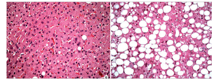
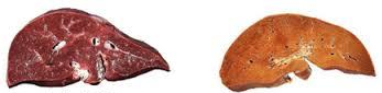
Fatty liver is a condition consisting of the accumulation of fat (triglycerides) in the form of vesicles within the livers hepatocytes.
Fatty liver is divided into two subcategories; alcoholic fatty liver disease ALFD and non-alcoholic fatty liver disease or NAFLD, though the clinical outcomes are nearly the same. The condition is also associated with other diseases that influence fat metabolism.
NAFLD is the accumulation of fatty deposits within liver cells that is not associated with alcohol use. Caused by conditions like metabolic disorder, glycogen storage diseases and acute fatty liver of pregnancy. Other causes are nutritional, like severe weight loss, malnutrition, high fat diets etc..
NAFLD is reversible, if left untreated, however there is potential for inflammation called non-alcoholic steatohepatits or NASH.
AFLD is cause by the chronic over consumption of alcohol, the liver metabolizes alcohol into toxic substance, acetalaldehyde that may damage hepatocytes, about 90% of chronic alcohol users develop fatty liver. IF left untreated AFLD can lead to alcohol hepatitis, fibrosis and ultimately cirrhosis.
Ultrasound
The liver echotexture is homogenous with echogenicity only slightly increased when compared to the renal cortex, and hypoechoic to the spleen.

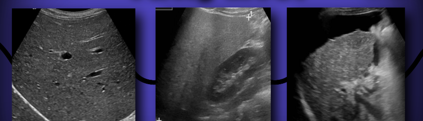
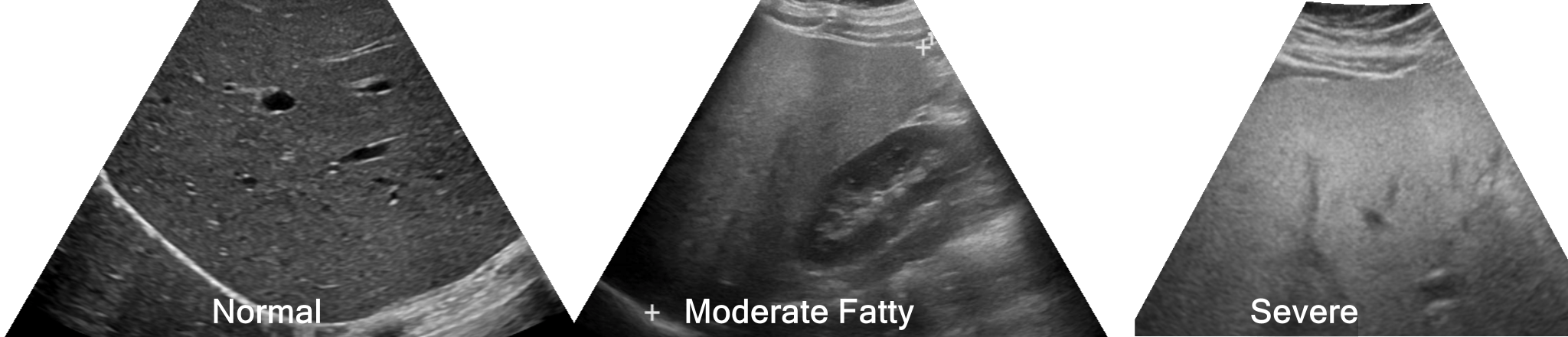
When there is fatty infiltration of the Liver the parenchyma becomes more echogenic (hyperechoic). Fatty liver can be described by ultrasound as mild, moderate and severe, though this is more a qualitative assessment, with mild fatty liver you’ll see increased echogenicity, and loss of the interface with the hepatic vein walls. With increasing severity you’ll lose the interface of the portal vein walls and biliary radicals to the liver parenchyma. Fat increases the attenuation of sound and in severe cases you can’t make out the posterior aspects of the right lobe of the liver, including the diaphragm and right kidney.
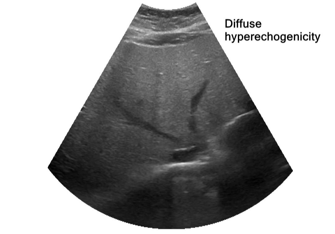
Patterns of fat accumulation can be diffuse, diffuse with sporadic areas of normal liver tissue (focal fatty sparing), focal or segmental infiltration can be seen as well, though less common.
Diffuse Fatty Infiltration
Focal Fatty Sparing

Focal Fatty Infiltration

The prevalence of fatty liver disease ranges from 25% to 35%. approximately 75% of obese people have fatty infiltration. It is the most common cause of abnormal liver function tests in the United States.
Patients are usually asymptomatic and present with “transaminitis” a term which is synonymous and used interchangeably with elevated liver enzymes and or transaminasemia, specifically aspartate aminotransferase [AST] and alanine aminotransferase [ALT] are elevated. You may also see an elevation of bilirubin and GGT Gamma-glutamyltransferase, though this is usually elevated in alcoholic liver disease it is not seen in up to 70% of patients who abuse alcohol.
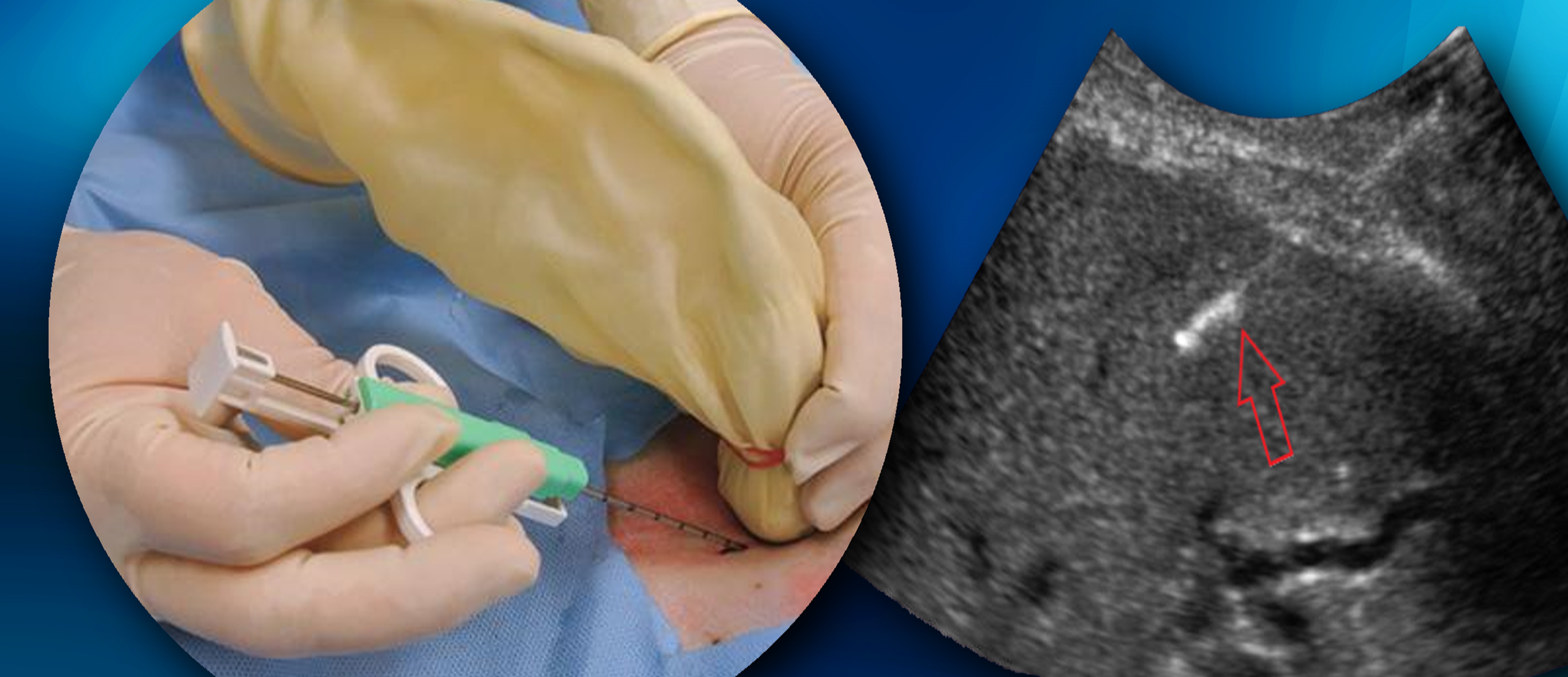
Liver Biopsy
Liver biopsy is the most sensitive and specific test in evaluating patients with AFLD. The biopsy is used to determine whether there is liver injury like fibrosis. The biopsy is done often under ultrasound guidance. The procedure is done with a team that includes a Sonographer, Physician, Registered Nurse and Laboratory Technologist. Using a sterile technique the procedure is often done in a supine position, the most adequate window is found and marked. The biopsy is taken through intercostal spaces, using the ultrasound to avoid hitting nearby structures like Kidney, Bowel, GB or Large vessels.
Sources:
http://emedicine.medscape.com/article/175472-overview#a1
http://ajpgi.physiology.org/content/290/5/G852
https://www.ncbi.nlm.nih.gov/pmc/articles/PMC3177433/
http://www.nature.com/nrgastro/journal/v9/n3/fig_tab/nrgastro.2011.273_F2.html

Great images and information. I wish I would have had this when I was in school many years ago. I’ll share this with our students at out clinical site. Thank you!
LikeLike
Oops “Our”
LikeLike
Excellent description…very informative…easy language.
LikeLike
You’re very welcome!
LikeLike
great reference/refresher for my students and staff.
LikeLike
😁 glad you found it useful!!!
LikeLike
Excellent. Great information. Very helpful. Will use it to help our ultrasound techs.
Tom Bowers MD
LikeLike
Thank you so much!
LikeLike
Awesome
LikeLike
Thanks everyone for the helpful information on the LIVER enzymes And all the other ways to reverse NAFLD.
LikeLike