


Upper extremity venous doppler is performed to rule out deep vein thrombosis (DVT). In order for an upper extremity venous thrombus to be considered a DVT the clot has to seen within the internal jugular (IJV), subclavian, axillary or brachial veins. One of the main concerns with DVT is that it can lead to Pulmonary Embolism, that’s when a piece of the clot is dislodged and travels to the pulmonary vasculature (embolus) which can be a life threatening event.
DVT’s are usually caused by stasis, immobility and hypercoagulable states, (Virchow’s triad).

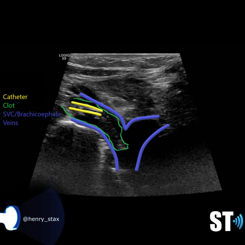
Upper extremity clots are commonly caused by venous lines, especially PICC lines (peripherally inserted central catheter). The basilic vein is the typical location for insertion of PICC lines, thrombosis can be seen in up to 70-80% according to some studies, especially the longer the line is in place.
Protocol
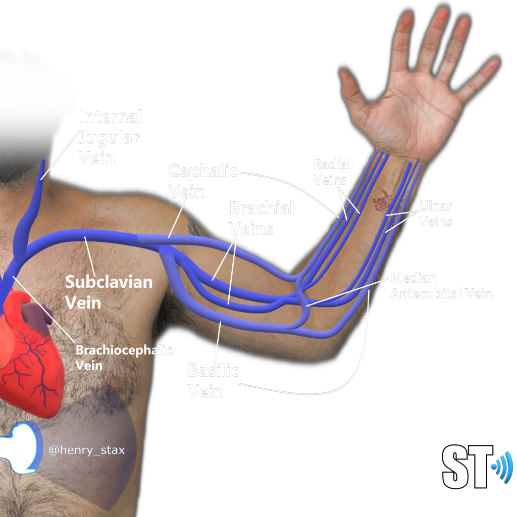
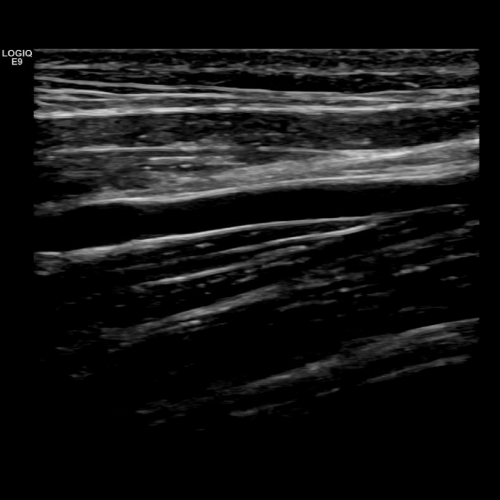
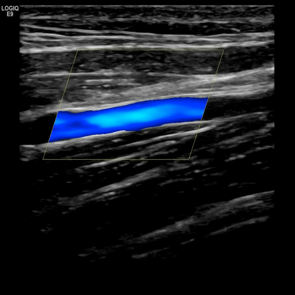
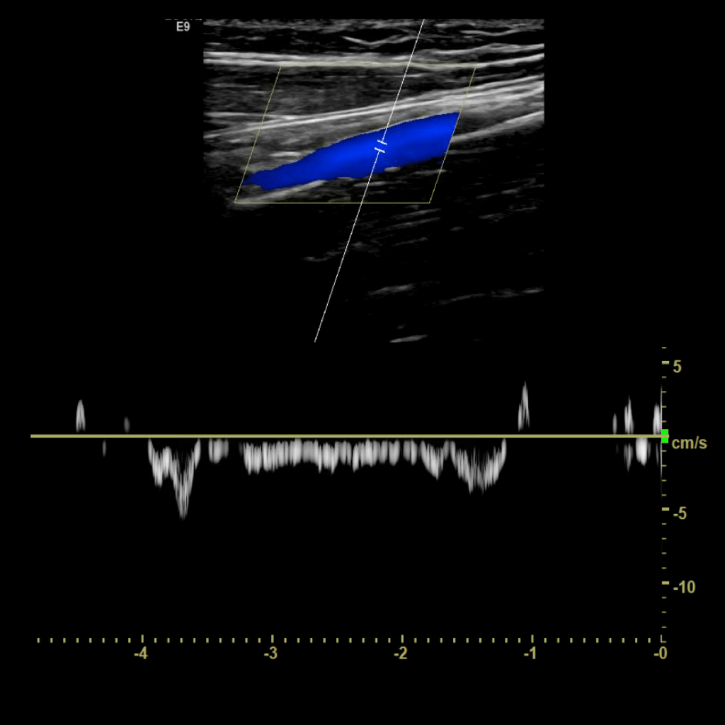
A typical scanning protocol includes transverse and sagittal images with and without color doppler and spectral wave analysis of the IJV, subclavian, axillary, brachial, basilic, cephalic, radial and ulnar veins. In my institution we include innominate and superior vena cava (SVC) in the vessels we interrogate.
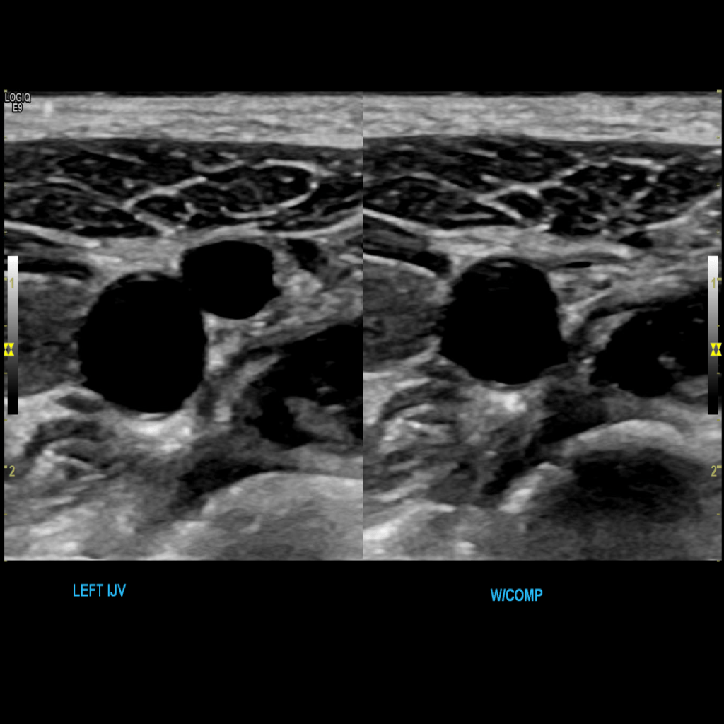
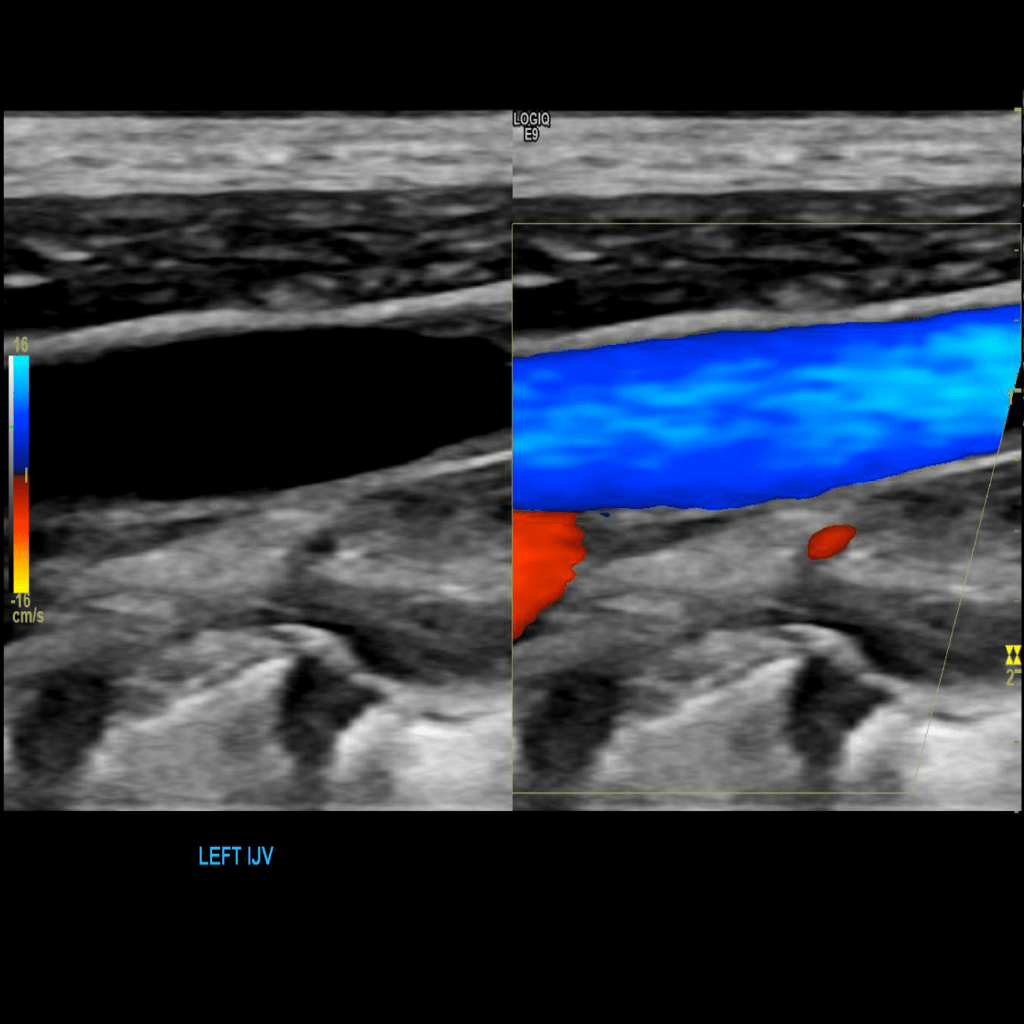
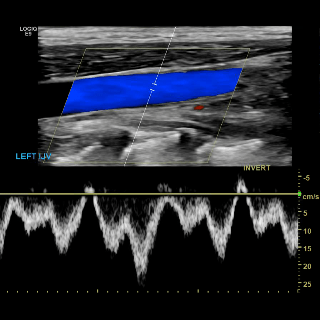
Scan the internal jugular vein in grey scale, compression, color doppler and spectral doppler
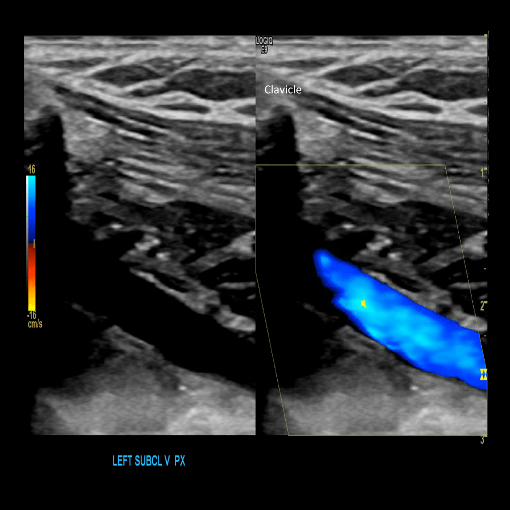
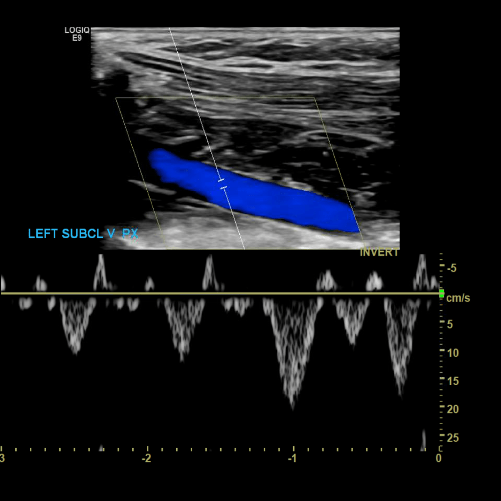
Scan the subclavian vein in grey scale, color doppler and spectral doppler
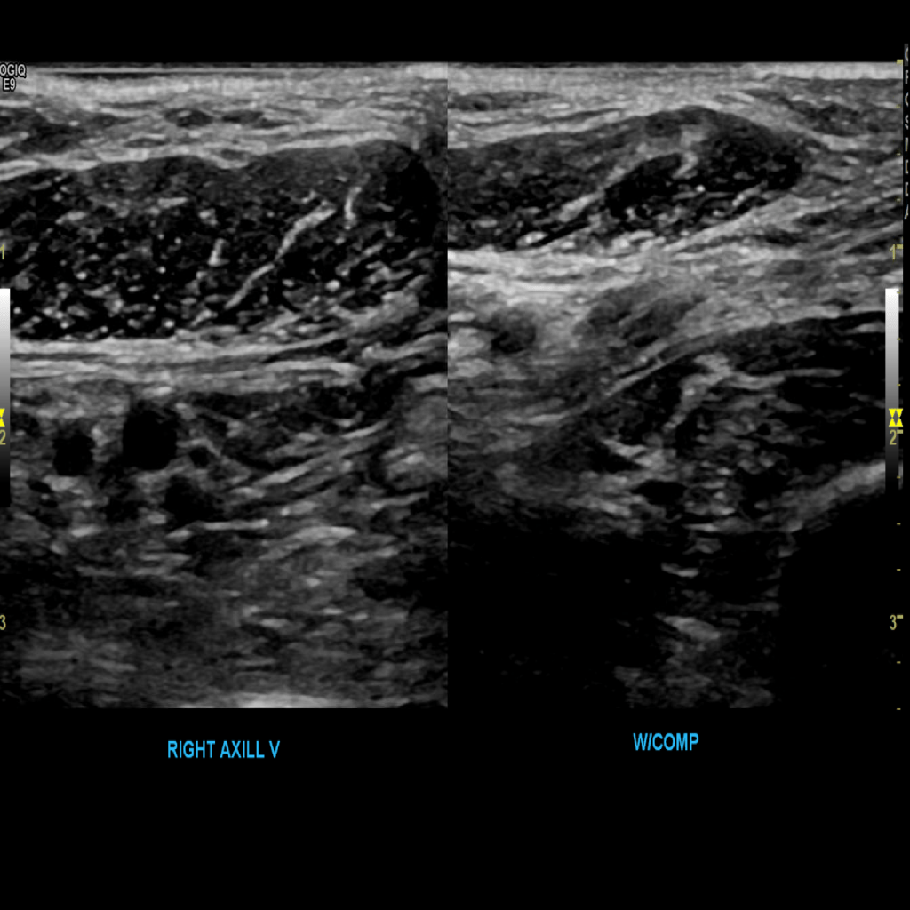
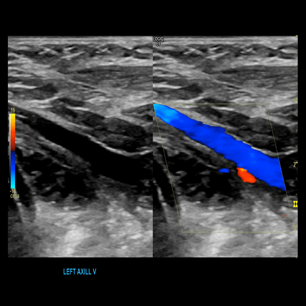
Scan the axillary vein in grey scale, compression, color doppler and spectral doppler

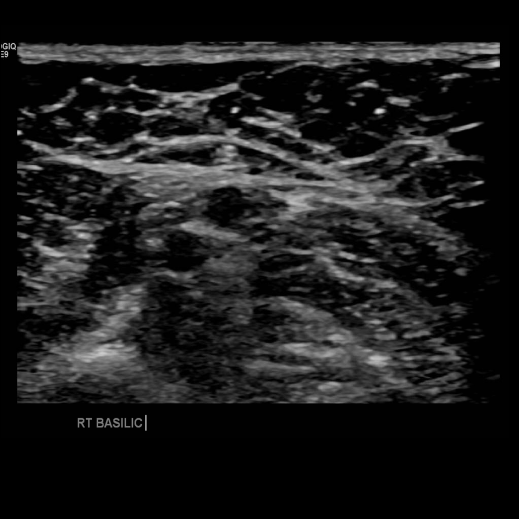
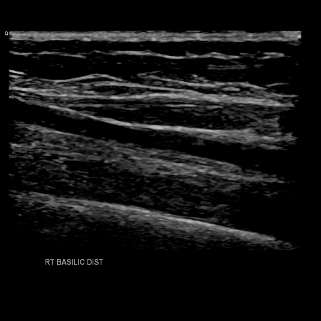
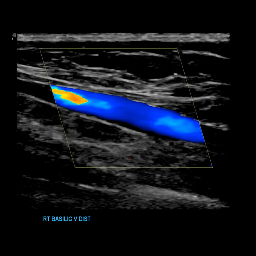
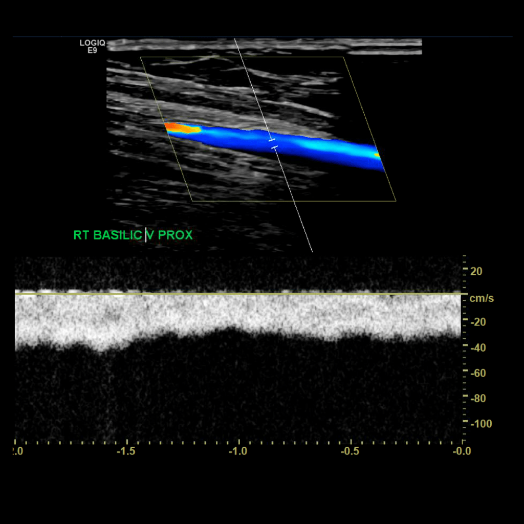
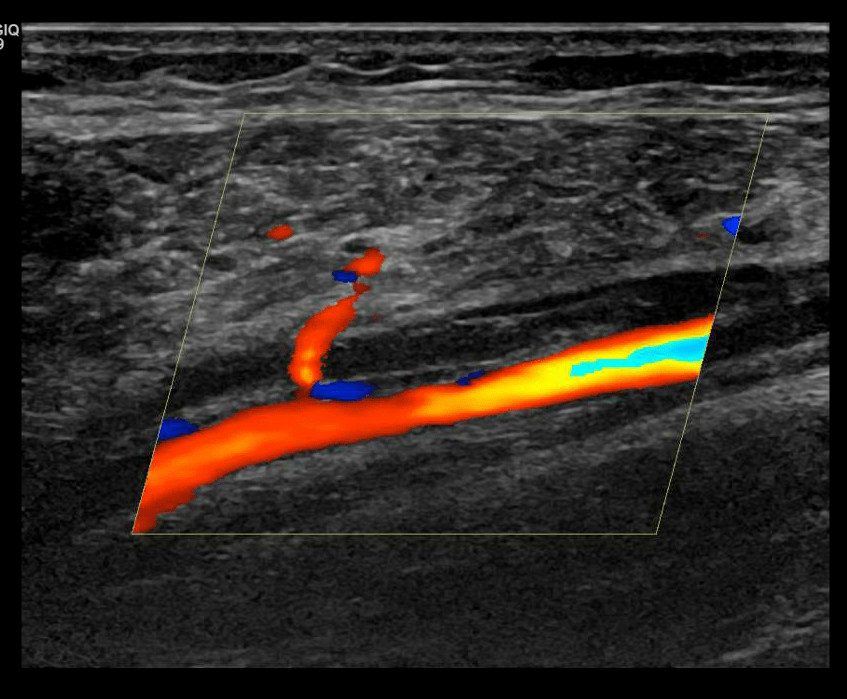
Scan the basilic vein in grey scale, compression, color doppler and spectral doppler
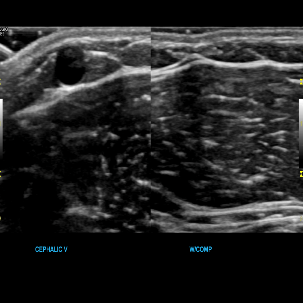
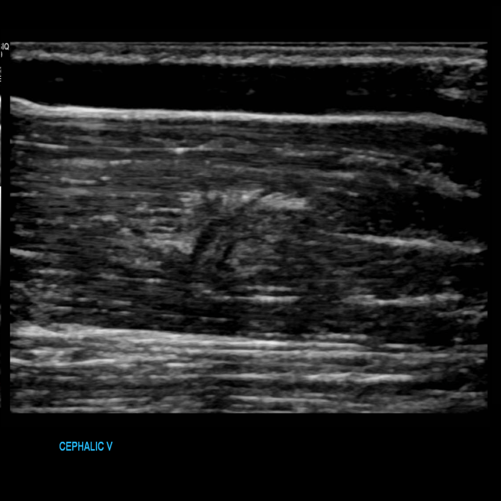
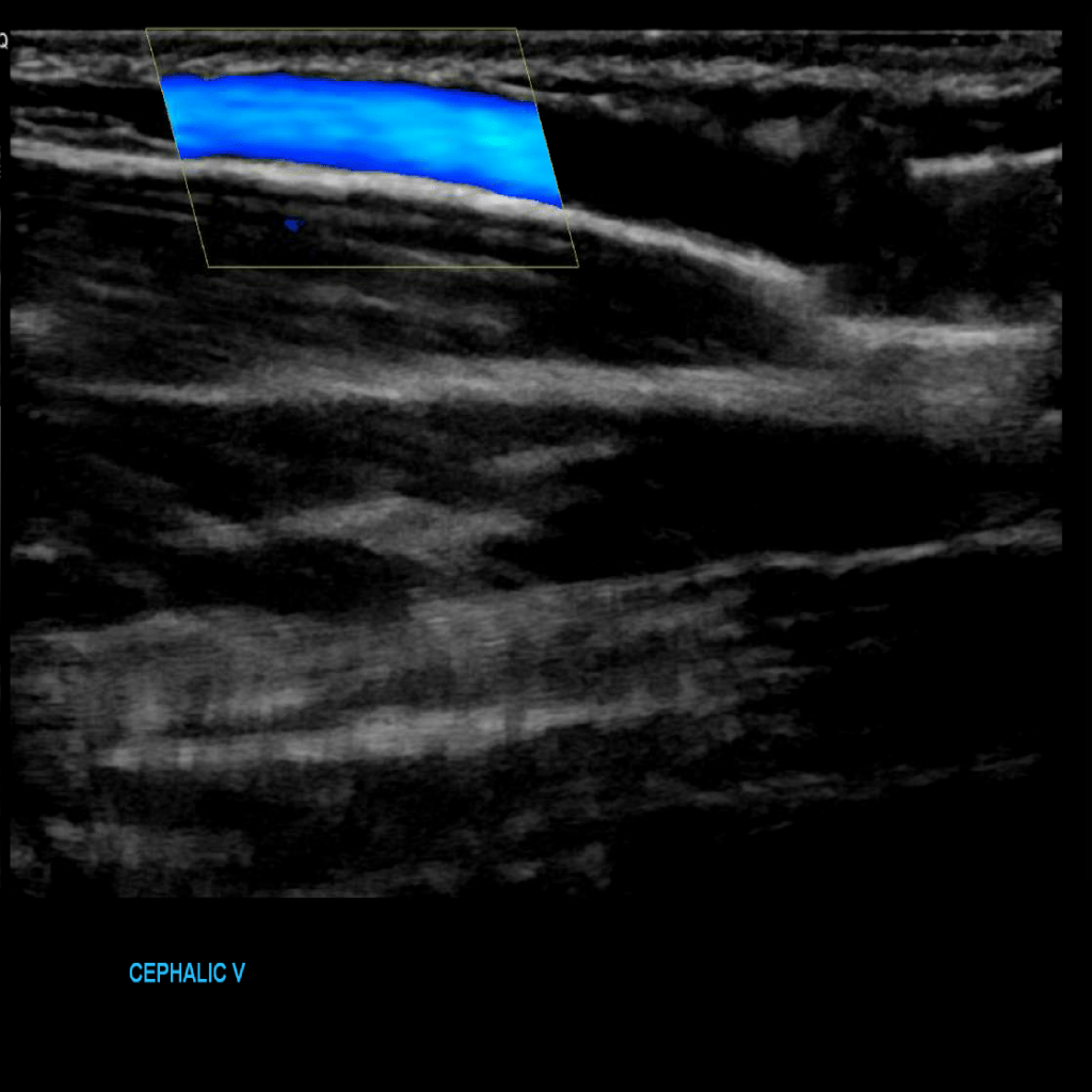
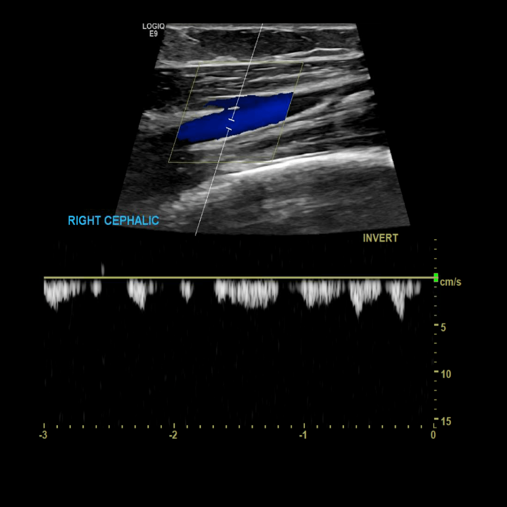
Scan the cephalic vein in grey scale, compression, color doppler and spectral doppler
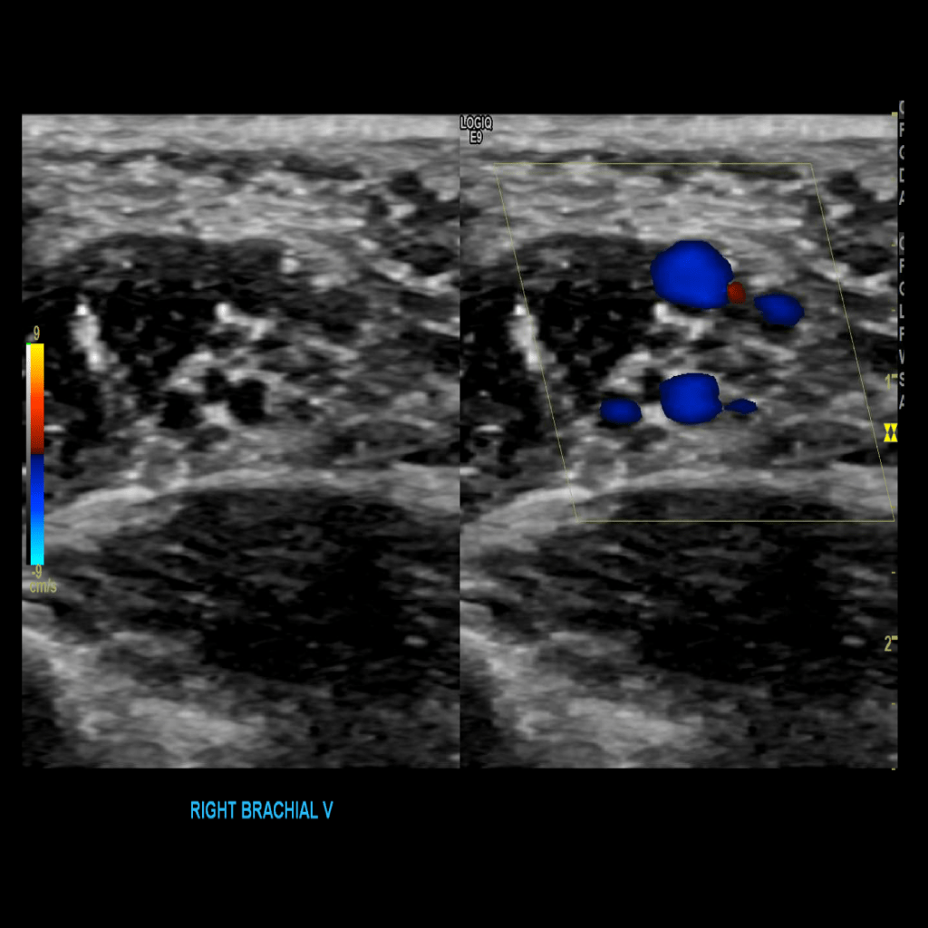
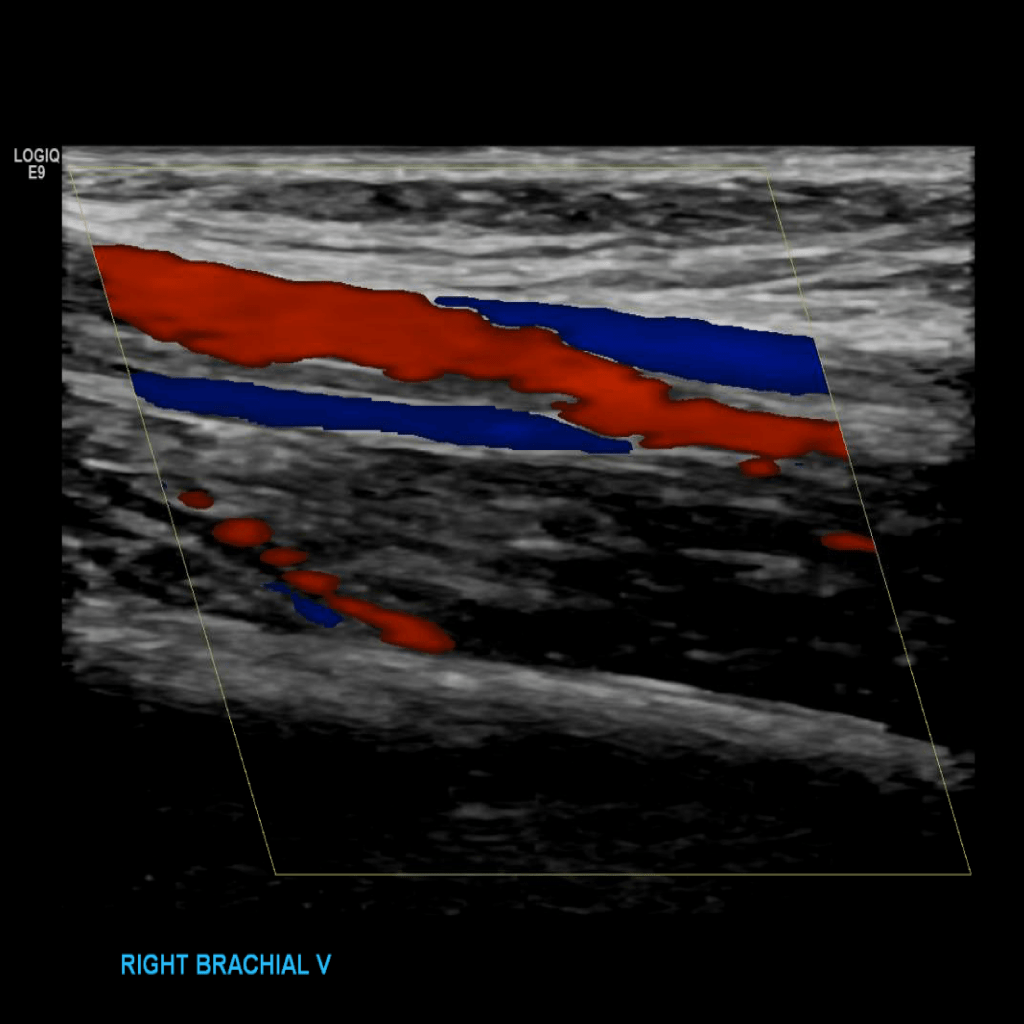
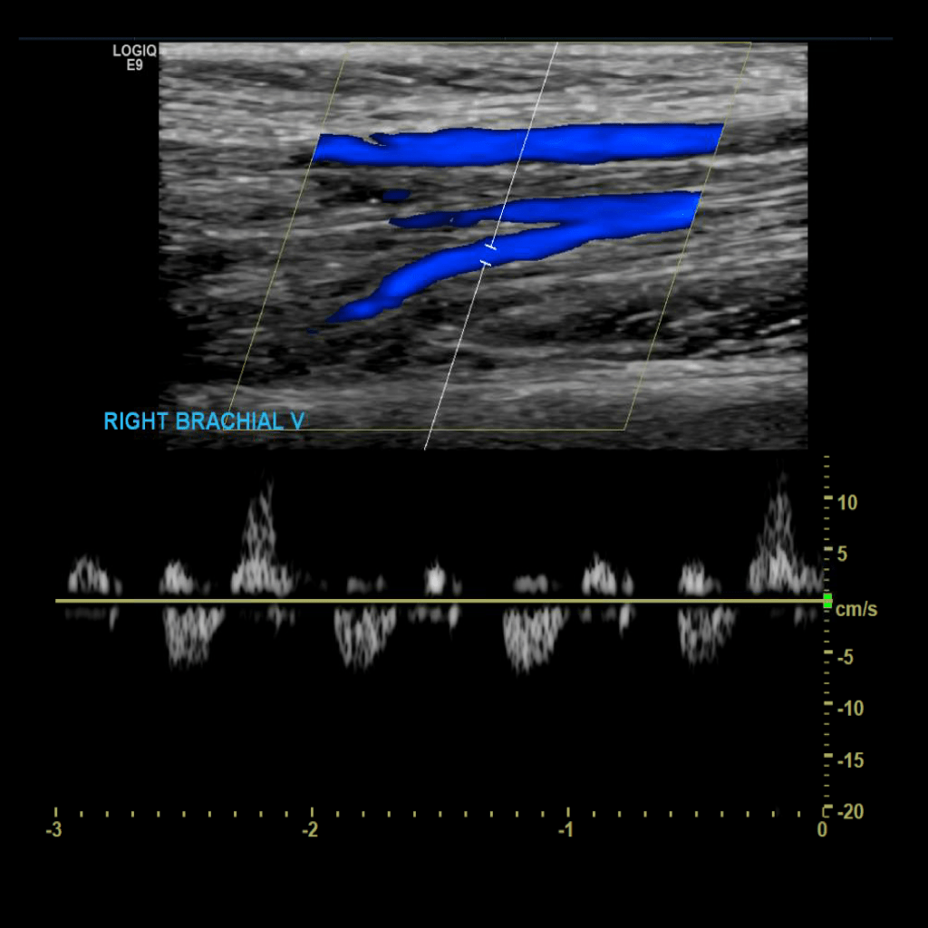
Scan the brachial veins in grey scale, compression, color doppler and spectral doppler
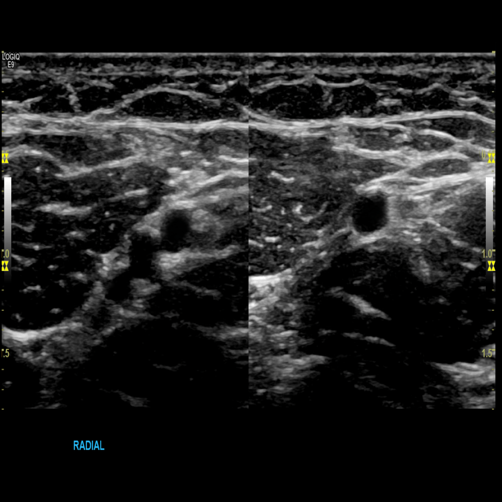
Scan the radial veins in grey scale, compression, color doppler and spectral doppler
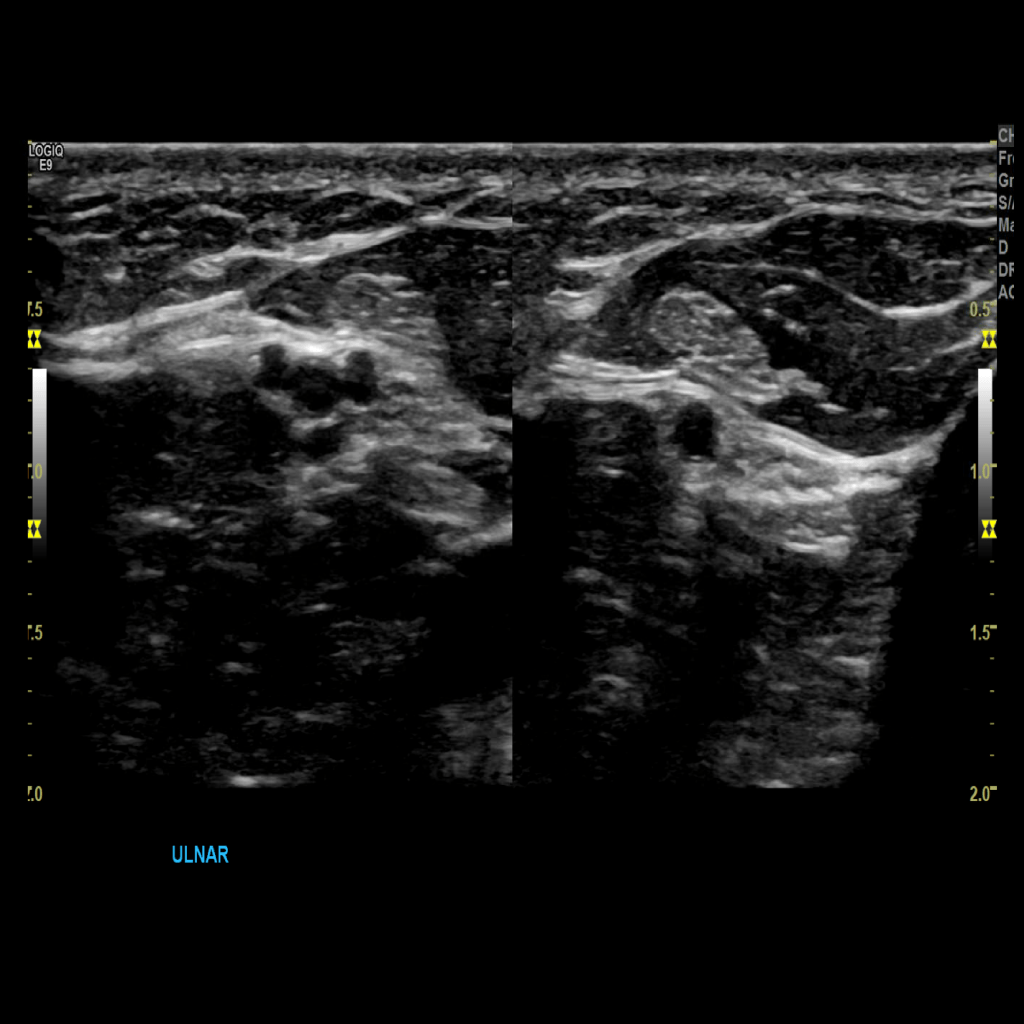
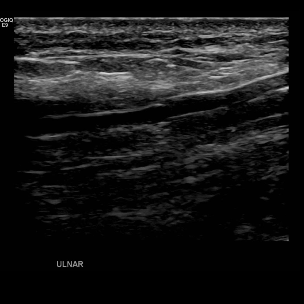
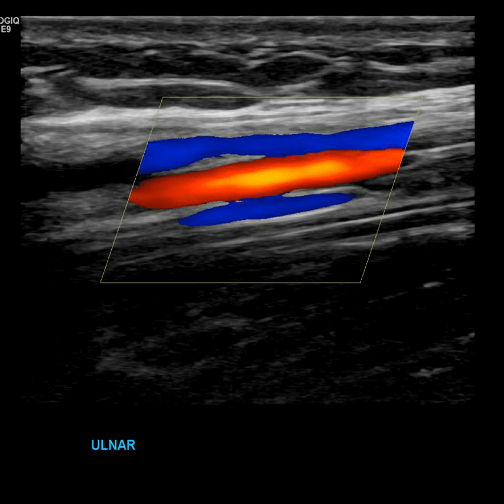
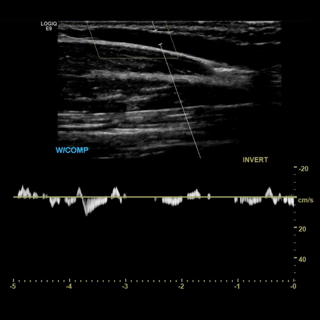
Scan the ulnar veins in grey scale, compression, color doppler and spectral doppler
Pathology
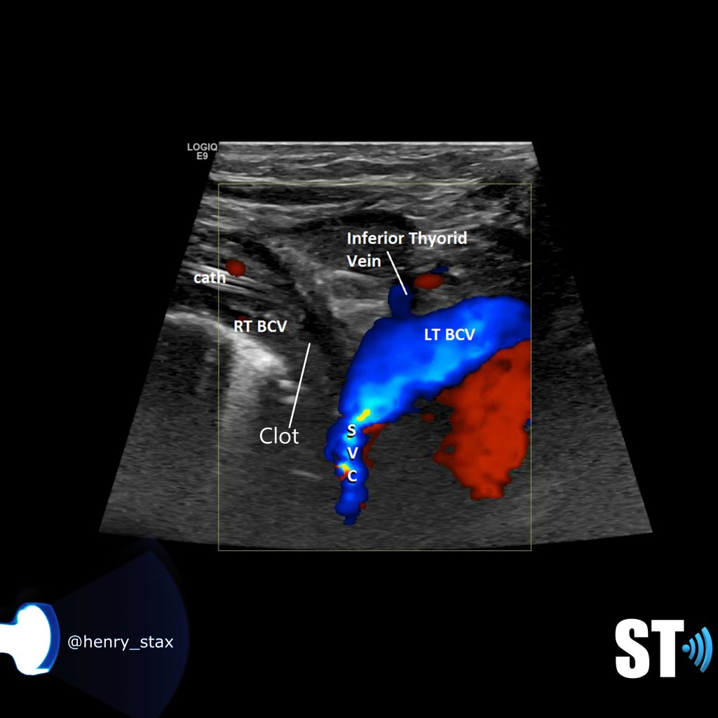
Thrombosis of the right innominate vessel with indwelling catheter
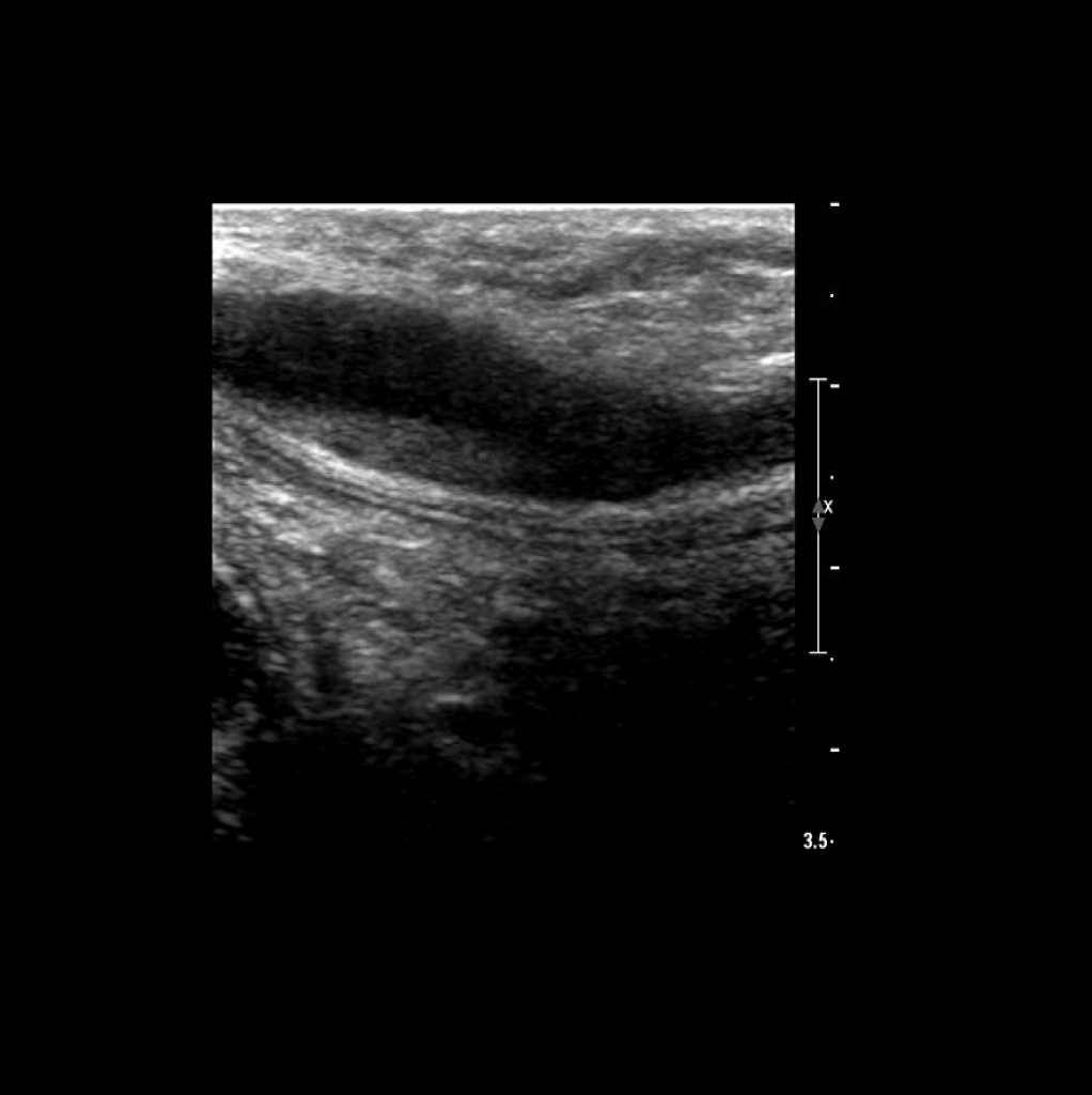
DVT of the subvlavian
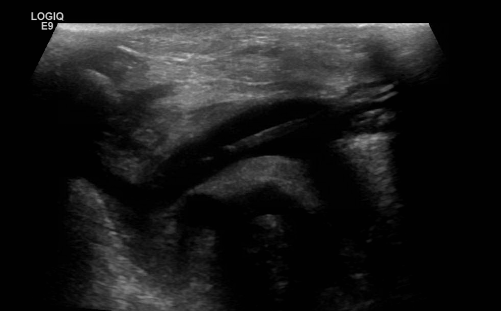
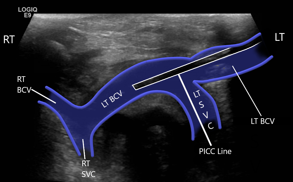
Duplicate SVC
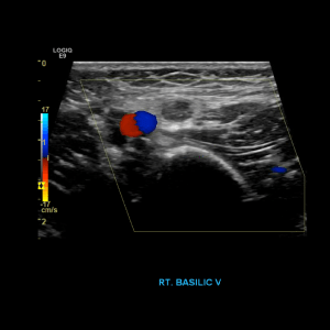
Thrombus of the right basilic vein
Brachial vein thrombosis
I have had only a handful of thrombosed upper extremity veins in the absence of a line. A couple of those patients were pitchers and had paget schroetter syndrome aka venous thoracic outlet syndrome.
Henry Suarez RDMS,RVT
Further reading:

Do you image more distally with arms?Thank you!
LikeLike
Depends on the institution, some places include the radial and ulnar arteries, others do not.
LikeLike
Can you give any tips on how to image the innominate and the supraclavicular subclavian? I have a difficult time visualizing these.
LikeLike
I would try scanning above the clavicle and angling down towards the heart. Use a sector probe for the smaller footprint and depth. And also try having the patient propping their head slightly up and towards the side you interested so the muscle is more relaxed..
LikeLike
As above I am having trouble scanning the subclavian. I tried scanning from on top of the clavicle down – but not great- then under the clavicle – not great – even if you did a video!!!!!!!!!?
LikeLike