

Musculoskeletal Sonography is becoming more utilized each day. We now have a certification via the ARDMS exclusively for MSK ultrasound. In no time it’ll be as common as gallbladders! It’s good to study and learn new techniques to make yourself marketable in the future of sonography and also cause learning is fun!
Routinely scanned structures are wrists, shoulders, knees, hips etc. Typically we are concerned with problems involving the muscles and tendons, looking for abnormalities including tears, ruptures, fluid collections and inflammation.
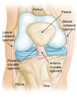
Shoulder

When scanning the shoulder you want to interrogate the biceps tendon, subscapularis, acromioclaivular joint, supraspinatus, glenhumeral joint and posterior labrum.
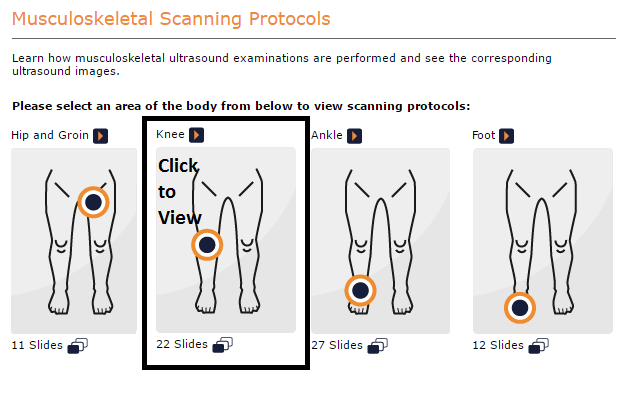
Shoulder Scanning Protocol (click image to view)
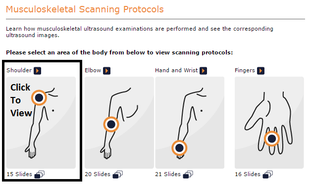

Labral Tear

Rotator Cuff Tear

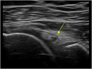
Supraspinatus Tear


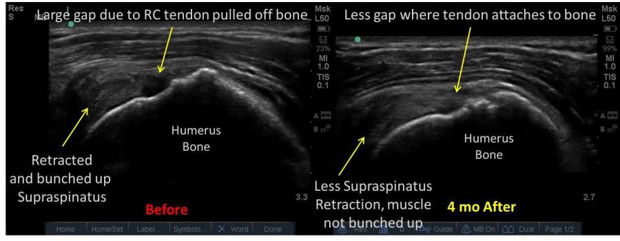
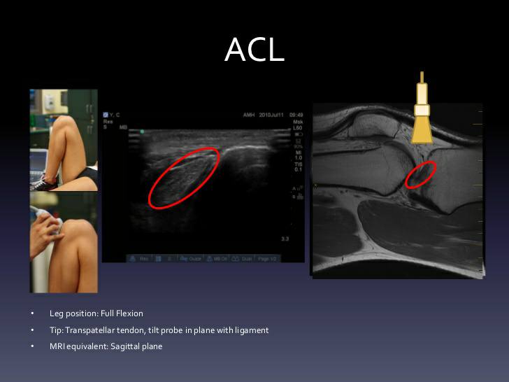
Anterior Cruciate Ligament
![]()
Knee Scanning Protocol (click image to view)

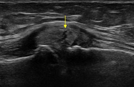
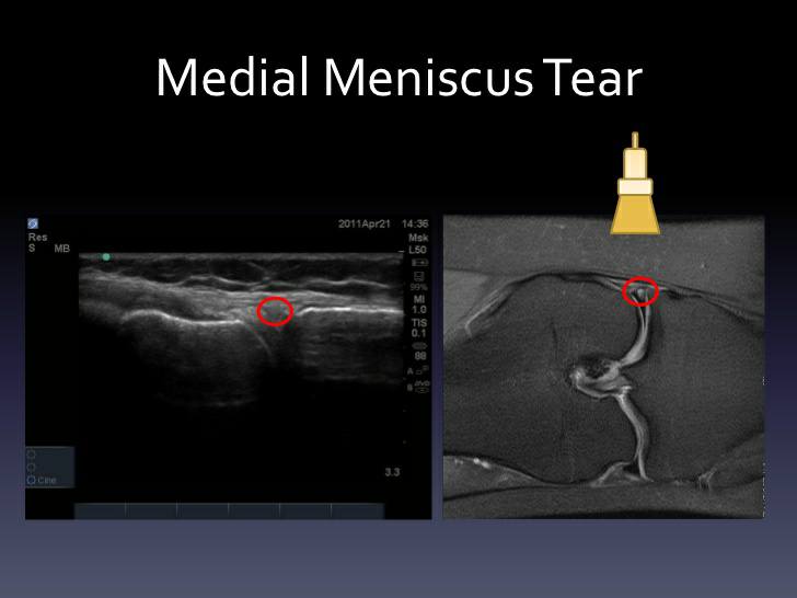
Meniscal Tears

Bulging meniscus with horizontal tear
These are just a few of the uses for MSK ultrasound, below I include a video that is an in depth analysis of MSK Sonography.
Infographics from mountsinai.org/sportsmedicine
