

 The gastrointestinal system (aka GI Tract, alimentary canal or digestive system) is a series of hollow tubes that begins in the mouth and ends in the anus. The primary function of the GI tract is to break down food into energy for the various metabolic processes involved with life.
Digestion begins in the mouth, teeth mechanically break down food into smaller pieces (mastication) which are coated with saliva (made by the salivary glands)
Submandibular glands


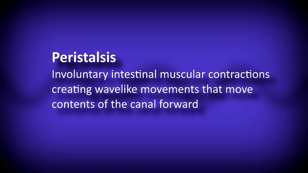
Saliva begins the chemical break down of food via salivary amylases and also coats the food making swallowing easier. Once food passes into the esophagus a series of wave like contractions propel food down its long journey, these waves are known as peristalsis and occur throughout.
Peristalsis
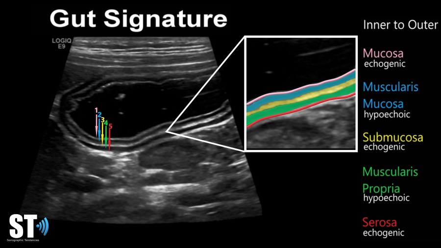
We use the “gut signature” to identify any gastric tissue in the abdomen. Bowel has 5 layers, made up of alternating hyperechoic and hypoechoic layers. These layers correspond to the histology of bowel. The layers are:
- mucosa: echogenic
- muscularis mucosa: hypoechoic
- submucosa: echogenic
- muscularis propria: hypoechoic
- serosa: echogenic
Once the bolus of food (referred to as chyme) reaches the end of the esophagus it encounters a sphincter known as the esophageal sphincter a muscular ring separating the esophagus from the stomach.
Esophagus


The stomach is a muscular pouch that further digests food mechanically though contractions and chemically with digestive enzymes and gastric acids.
Pathology
- Not routinely scanned sonographically
Stomach


At the end of the stomach you’ll find another sphincter the pylorus. After food passes through here it enters the first part of the intestines, the duodenum.
Pathology
- Gastric Tumors (Teratoma <1 %, Carcinoma 0.05%)
- Bezoar
- Hypertrophic Pyloric Stenosis
Pylorus


Small Intestine
The small intestine is much longer than the large intestine with a length varying from 9ft to 34.4ft. Food entering the small intestine mixes with secretions rich in sodium bicarbonate that neutralizes the stomach acids and allows for nutrients to be absorbed.
The small intestines are made up the duodenum, the jejunum and ileum.


Most of the nutrients absorbed in the small intestine are absorbed in the jejunum. Exceptions to this include iron, bile salts, water, lipids, fructose, and vitamin B12. The gallbladder releases bile and the pancreas releases the digestive enzymes amylase and lipase, they enter through the ampula of Vater in the duodenum.
Pathology
- Small Bowel Intussusception
- Inflammatory Bowel Disease
- Necrotizing Enterocolitis
- Henoch-Schönlein Purpura
- Meckel’s Diverticulum
- Duplication Cyst
Large Intestine
Right after the terminal ileum begins the first part of the colon, the Cecum. Food entering the cecum is controlled by the ileocecal valve. Off the inferior medial portion of the cecum arises the appendix, a tubular, blind ending continuation. The large intestine is divided in ascending, transverse, descending, sigmoid and finally rectum and anus. The large intestine is wider than the small intestine and its length is usually about 5 feet. The large intestine is mainly used for the passage of waste and re-absorption of water, though healthy bacteria also live here that make up the gut microbiome. The surface of the large intestine is marked by outpouchings and grooves called haustra, which happen due to a tight muscular band that runs the length of the colon called tinea coli. This acts similar to the elastic of hair scrunchies.


Pathology
- Intussusception Ileocolic – 77%
- Inflammatory Bowel Disease (i.e. Crohn’s, UC, Infectious)
- Diverticulosis/itis
- Appendicitis
Differentiating small and large bowel
We use anatomical location and differences to differentiate between large intestine and small intestine. Small intestine is quite disorganized and appears as multiple loops of free bowel usually with fluid, air or both, the wall are normally quite thin. Large intestine on the other hand has the haustral fold and is broader as previously mentioned.
Appendix
The appendix is a finger like projection off the base of the cecum which can be fluid filled, though sometimes fecal matter and air can be seen withing the lumen. It is normally under 0.6 cm and is compressible.


Pathology
- Appendicitis
- Mucocele of the appendix
- Appendiceal carcinoma

Great lectures! The videos are very educational and easy to follow!
LikeLike
😌 glad you like it!
LikeLike