

Here is a quick video explaining the protocol.
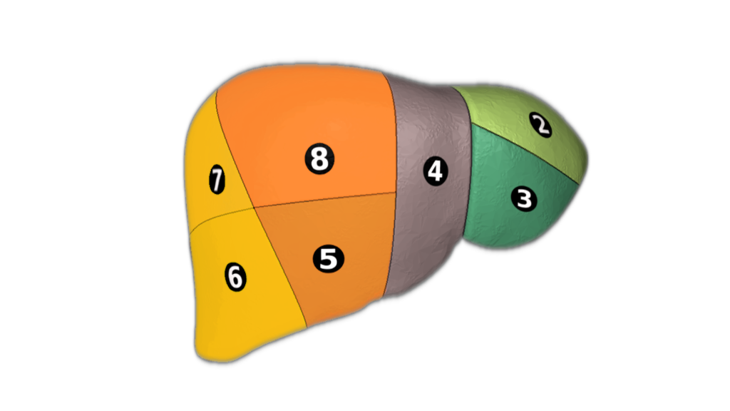
Liver Protocol basics. The liver is the largest internal organ, the right lobe is 5-6 times larger than the left. Normally measures about 15 cm at mid clavicular line in adults, there is much variation in pediatric populations by age and size. The liver is covered by a layer of connective tissue referred to as Glisson’s capsule. The liver is normally more echogenic then the renal cortex. Order of echogenicity highest to lowest renal sinus> pancreas>liver>spleen>renal cortex >renal medullary pyramids The liver is drained by the hepatic veins of which there is usually 3, Left, Middle and Right. They connect to the IVC which drain into the right atrium. The liver is fed oxygenated blood by the hepatic artery and portal vein. Here is the basic sonographic protocol for scanning the liver.

Sagittal Views


Transverse Views


Your first image is of the lateral left lobe including the in a longitudinal plane.
Moving medial you’ll encounter the aorta. Be sure to include the celiac and superior mesenteric arteries
The following image should include the caudate lobe and ligamentum venosum followed by the inferior vena cava.
Next up is the gallbladder you can scan subcostally, having the patient take a deep breath and holding it or through the ribs. From here on you’ll be interrogating the right lobe of the liver. Here you can encounter the portal triad (portal vein, common bile duct and hepatic artery) and the gallbladder
Moving laterally again through either between or under the ribs at the midclavicular line you can get the liver in its longest span and measure in a craniocaudal fashion.
Further lateral you’ll encounter the right kidney. The kidney should be less echogenic to the liver. You can also assess the diaphragm, check for pelural effusion or free fluid in Morrison’s pouch. Be sure to image the dome of the liver.
Moving on to the transverse views begin again in the left lobe angling superior. You can see segments I, II and II of the liver along with the left portal vein.
Angling caudally you can see more of segment II and segment I i.e. caudate lobe. Also imaged here is the intrahepatic IVC and aorta .
Angling further down you’ll encounter the pancreas and mesenteric vasculature. In very young or slender patients you may be able to see the kidneys on either side of the vertebrae.
Moving back up and angling slightly obliquely you can see the hepatic veins, a view know as the “playboy bunny” sign.
Next is the right and left portal veins as well as the IVC.
Transverse right lobe.

EXCELLENT POST
LikeLike
Thank you!
LikeLike
perfect
LikeLike
Thanks!
LikeLike
ottimo
LikeLike
thanks a lot for the simpliest and clearest explanation
LikeLike
Soy una principiante, y he leído varios recursos para orientarme y esta información es la más didáctica y entendible que he encontrado… gracias!
LikeLike