

Intracranial Hemorrhage of Prematurity
Intraventricular hemorrhages aka Germinal Matrix/Subependymal bleeds are believed to happen due to poor autoregulation of premature infants, sudden changes in pressure or oxygen saturation can cause sudden reperfusion which ruptures of the tiny delicate vessels within the germinal matrix
Etiologic Factors

Prevalence
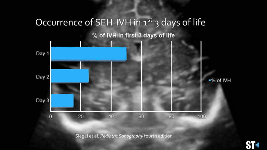
Most of the Bleeds occur early
Day 1 50%
Day 2 25%
Day 3 15%
Day 4 10%
95% of all bleeds occur by day 9
The intraventricular hemorrhage is typically categorized into 4 grades:


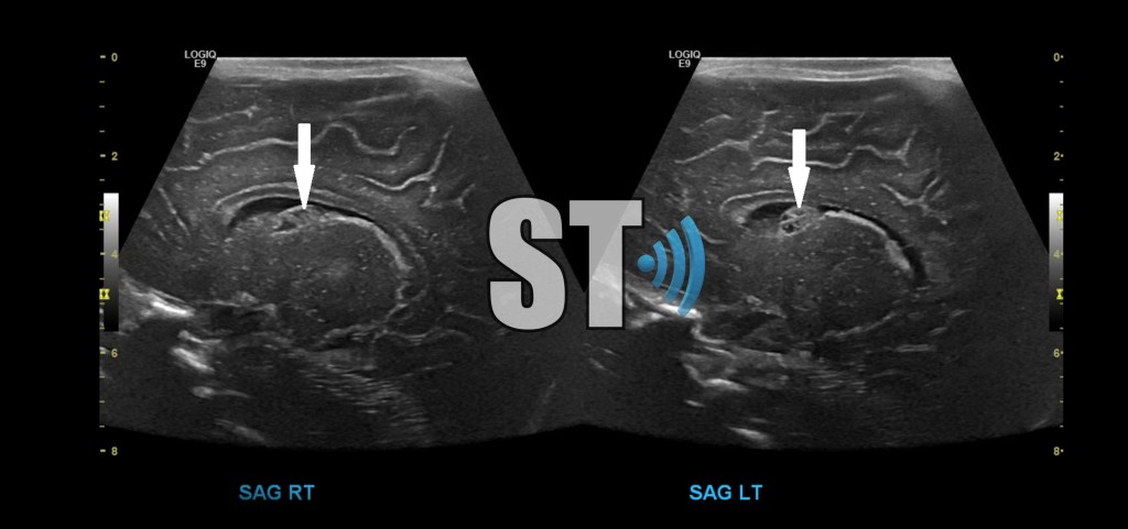
Grade I IVH
Bilateral grade I IVH, close follow up is recommended to ensure complete resolution.
Grade II IVH
Grade III
Coronal and Sagittal images of a premature neonate with bilateral grade III IVH, notice the lack of gyri and sulci indicative of a extreme prematurity.

Grade IV IVH
Initial images of a grade IV intraventricular hemorrhage (upper image) and 2 month follow up showing a large cystic cavity where the hemorrhage was (cystic encephalomalcia).

A subependymal (grade I) bleed will eventually liquify and undergo cystic degeneration. 80% extend into the Intraventricular region (grade II). 15% of bleeds will also develop in the intraparenchymal hemorrhages (grade IV). It was thought that intraprenchymal bleeds started in the germinal matrix but now it is believed that they occur from venous infarcts.

Sequlae of ICH
Cysts (porencephalic cysts and periventicular leukomalacia)
Encephalomalacia (necrosis of white matter with liquefaction and cavitation) ventricular dilatation.
- 70% infants with IVH are mild and resolve
- 15% will be severe
- 15% or less require shunts
Hypoxic-ischemic encephalopathy is one of the most common causes of cerebral palsy and other severe neurological deficits in children, occurring in 2-9 of every 1000 live births.
“State-of-the-Art Cranial Sonography: Part 1, Modern Techniques and Image Interpretation : American Journal of Roentgenology: Vol. 196, No. 5 (AJR).” State-of-the-Art Cranial Sonography: Part 1, Modern Techniques and Image Interpretation : American Journal of Roentgenology: Vol. 196, No. 5 (AJR). N.p., n.d. Web. 07 Nov. 2016.
Ultrasound Q. 2002 Jun;18(2):89-114.
Intracranial neonatal neurosonography: an update.
Benson JE1, Bishop MR, Cohen HL.
Radic, Julia A. E., et al. “Outcomes of Intraventricular Hemorrhage and Posthemorrhagic Hydrocephalus in a Population-Based Cohort of Very Preterm Infants Born to Residents of Nova Scotia from 1993 to 2010.” Journal of Neurosurgery: Pediatrics, vol. 15, no. 6, 1 June 2015, pp. 580–588

Excellent presentation, very helpful indeed
regards Florence >
LikeLike
😌
LikeLike
This presentation is excellent and very helpful especially for teaching purposes
LikeLike
I’m glad you liked it thanks for leaving a comment!!
LikeLike
[…] Intracranial Hemorrhage of Prematurity […]
LikeLike