

Molar Pregnancy – Hydatidiform Mole
Tweet
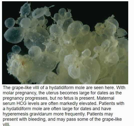
Hydatidiform Mole aka Molar pregnancy is cause by implantation and proliferation of a non-viable fertilized ovum. Characteristics are that of throphoblastic proliferation and Vesicular swelling of the chorionic villi.
Sonographically you’ll see an enlarged , round uterus with many cystic spaces classically referred to as “bunch of grapes” 🍇.
There are two types of molar pregnancies
Complete mole has a diploid genotype (46XX, 46 XY) it lacks fetal tissue. Complete hydatidiform moles have a 2–4% risk of developing into choriocarcinoma in Western countries and 10–15% in Eastern countries and a 15% risk of becoming an invasive mole.
hCg usually >100,000
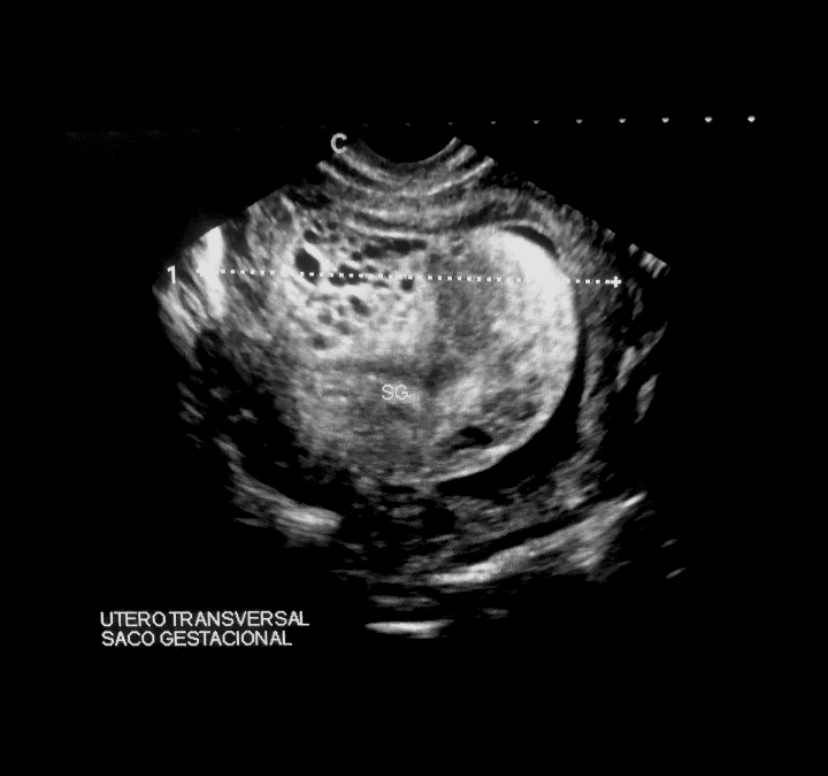
Case 1
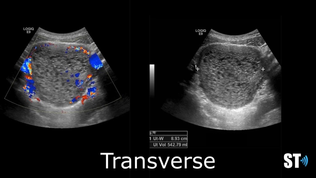
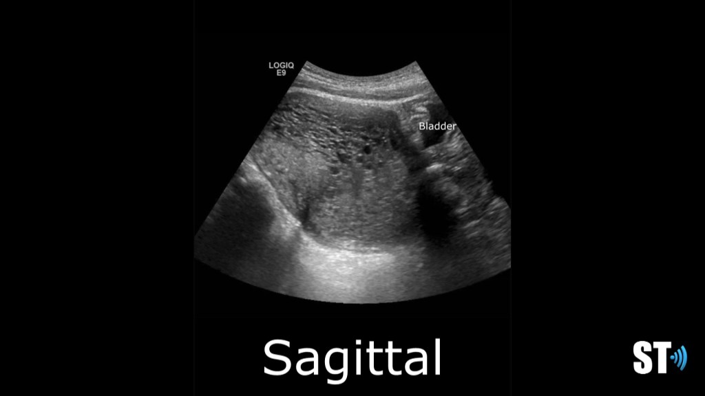
Complete mole transverse and sagittal images showing an enlarged, round uterus with a complex multi cystic endometrial complex. Hcg was 25,344
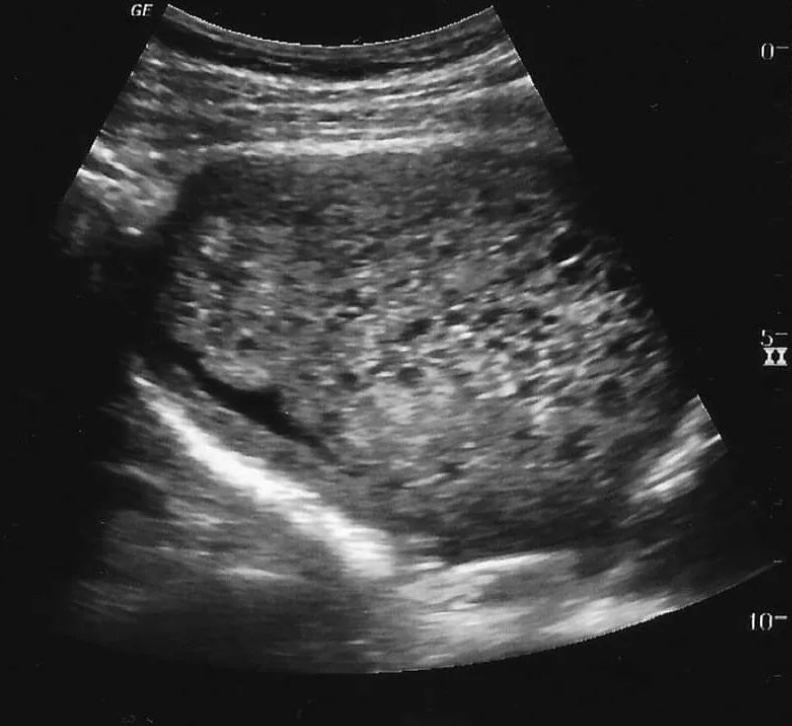
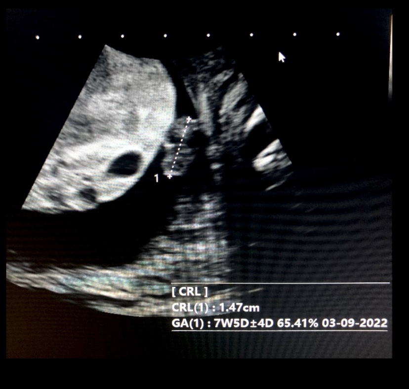
Case 2


Incomplete moles have Triploid genotype (69XXY, 69XXX) usually contains fetal/embryonic tissue/cells. hCg usually <100,000
<5% become invasive
Case 3


References:
- “Gestational Trophoblastic Disease”. American Cancer Society. 14 April 2011.
- Cotran RS, Kumar V, Fausto N, Nelso F, Robbins SL, Abbas AK (2005). Robbins and Cotran pathologic basis of disease (7th ed.). St. Louis, Mo: Elsevier Saunders. p. 1110. ISBN 0-7216-0187-1.
- Kumar V, ed. (2010). Pathologic Basis of Disease (8th ed.). Saunders Elsevier. pp. 1057–1058. ISBN 978-1-4377-0792-2.
- Di Cintio E, Parazzini F, Rosa C, Chatenoud L, Benzi G (November 1997). “The epidemiology of gestational trophoblastic disease”. General & Diagnostic Pathology. 143 (2–3): 103–8. PMID 9443567.
