

Case
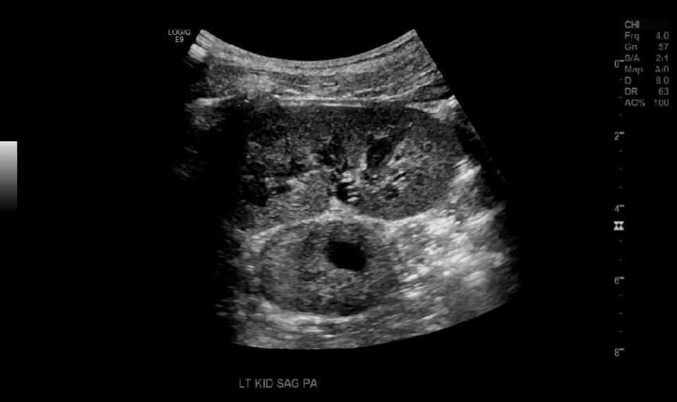
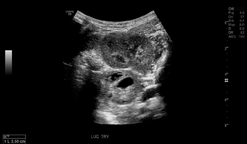
An adolescent patient presented for ultrasound with a history of tachycardia and diaphoresis. On ultrasound examination a heterogenous mass was seen medial to the left renal pelvis which was determined to be extra adrenal pheochromocytoma after further investigation.

Pheochromocytoma’s are rare adrenal tumors that arise from the medulla (chromaffin cell tumors) derived from the neural crest. They are associated with catecholamine secretion, primarily epinephrine and norepinephrine, and rarely dopamine. Large tumors secrete both epinephrine and norepinephrine
Potentially fatal, but with treatment carries a good prognosis.
Extra-adrenal pheochromocytomas may occur in the abdomen, thorax, urinary bladder, and neck and in association with the 9th and 10th cranial nerves.
The first histologically proven case of pheochromocytoma was
diagnosed by Felix Fraenkel at the University of Freiburg, Germany. [1]
Pheochromocytoma’s are rare tumors, with an annual incidence of 2 to 9.1 per 1 million adults and may correspond up to 60% of all adrenal incidentalomas [1]
Potentially inherited as an autosomal recessive trait.
Associated complications
- Cerebrovascular accident
- Retinopathy
- Irreversible kidney damage
- Acute pulmonary edema
- Cholelithiasis
- Cardiac arrhythmias
- Heart failure
Symptoms
Sustained or paroxysmal hypertension associated with headaches, sweating, or palpitations, occurs in 95% of patients, but at least 5% are normotensive. [2]
Patients often present with unpredictable episodes of hypertensive crisis paroxysmal symptoms suggesting a seizure disorder or anxiety attack. Hypertension that responds poorly to conventional treatment (<0.05% of hypertensives have a pheochromocytoma) [2]
Hypotension or shock after surgery or diagnostic procedures, headache, diaphoresis, palpitations, tremor
and nausea. Seeing as these symptoms can be seen in many conditions makes diagnosing pheochromocytomas difficult, and the list of differential diagnoses vast.
Mnemonic
There is a helpful mnemonic for pheochromocytomas, the 5 P’s.
- Pain (headache
- Pressure (hypertension)
- Palpitations
- Perspiration
- Pallor
Less than 20,000 cases per year in the US and only 10% of these are seen outside of the adrenal gland…
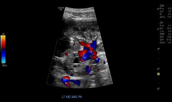
CT and MRI are the imaging of choice. On ultrasound, pheochromocytomas have a variable appearance ranging from solid (75% in one case series) to mixed cystic and solid to cystic. [3] IN our case the lesion was located near the left renal hilum and had cystic and solid components and some vascularity on color doppler.
Ultrasound


Treatment
The management of pheochromocytoma requires a multidisciplinary team. The only known curative treatment is surgical. In cases of surgery patients will undergo a thorough preoperative assessment including; patient and family history, complete blood count, metabolic profile, plasma metanephrines, ECG and cardiac ultrasound (to check for cardiac compromise). [1] Surgery for pheochromocytoma is considered a challenge due to the unpredictable release of catecholamines and rich blood supply. In one meta analysis involving 626 patients it was determined that laparoscopic removal of pheochromocytoma’s was safe and effective when compared to open surgery, with the caveat that there was no postoperative difference in blood pressure control, rates of severe complications, postoperative hypotension or cardiovascular disease between open and laparoscopic adrenelectomy. [4]
Bibliography
- Farrugia FA, Charalampopoulos A. Pheochromocytoma. Endocr Regul. 2019 Jul 1;53(3):191-212. doi: 10.2478/enr-2019-0020. PMID: 31517632.
- Manger WM, Gifford RW. Pheochromocytoma. J Clin Hypertens (Greenwich). 2002 Jan-Feb;4(1):62-72. doi: 10.1111/j.1524-6175.2002.01452.x. PMID: 11821644; PMCID: PMC8099329.
- Katherine Leung, Michael Stamm, Asim Raja, and Gavin Low, Pheochromocytoma: The Range of Appearances on Ultrasound, CT, MRI, and Functional Imaging, American Journal of Roentgenology 2013 200:2, 370-378
- Fu SQ, Wang SY, Chen Q, Liu YT, Li ZL, Sun T. Laparoscopic versus open surgery for pheochromocytoma: a meta-analysis. BMC Surg. 2020 Jul 25;20(1):167. doi: 10.1186/s12893-020-00824-6. PMID: 32711496; PMCID: PMC7382066.
