

The small organ that can cause big trouble.
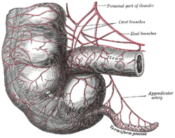
The vermiform (worm like) appendix is a finger-like projection off of the base of the cecum, it can become inflamed and require surgical removal.
Appendicitis
Symptoms:
- mid abdomen to RLQ pain
- rebound tenderness
- nausea, diarrhea or vomiting
- elevated white blood cells (not always)
- pain when walking or lifting right leg)
Scanning:
Use a linear probe 7-9 mHz. Try others if difficult to image patient.
Patients are scanned in supine and left lateral decubitus. Start at the RUQ image the Liver, RT Kidney and Gallbladder, sometimes the appendix may be tucked up there. Ask patient where maximum point of pain is. The normal location of the appendix is usually McBurney’s point (halfway between anterior iliac spine and umbilicus)

Use guided compression of the ascending colon down to the cecum
appendix can vary in location and be behind the cecum (retrocecal), deep in the pelvis, behind the bladder, draped over the iliac vessels, in the RUQ, midline near spine.


Normal Appendix:
-< 0.6 cm
-compressible
-may contain air bubbles or fluid
-is smooth does not contain haustra (sacular folds of the colon)
Normal Appendix

RUQ Appendix below RT Kidney anterior to psoas muscle

RUQ Appendix

RUQ Appendix
Positive Appendix:
-> 0.6 cm
-hyperemic (increased blood flow on doppler indicative of inflammation)
-non-compressible
-free fluid (may be present in normal, if complex or debris filled in the presence of appendicitis consider rupture
Positive Appendiceal Ultrasound

LArge appendix with appendicolith

Appendicitis with hyperemia

B-Flow hyperemia

transverse appendix with free fluid
Ruptured Appendicitis

Longitudinal enlarges appendix with fat stranding

Complex collection near transverse appendix

collection extends to behind bladder

collection
Henry Suarez RDMS, RVT
Image sources
Further Reading:

Very good. Thank you.
LikeLike
how hard is it to find? I’m new at this and I have no idea. I just read your blog and I love. Thank you
LikeLike
At first kinda difficult but when you’re done a bunch it’s pretty simple, though sometimes I’m unable to find one. Like in a given night I’ll ldo like 6, and sometimes I won’t find one
LikeLike