

Proximal Aorta
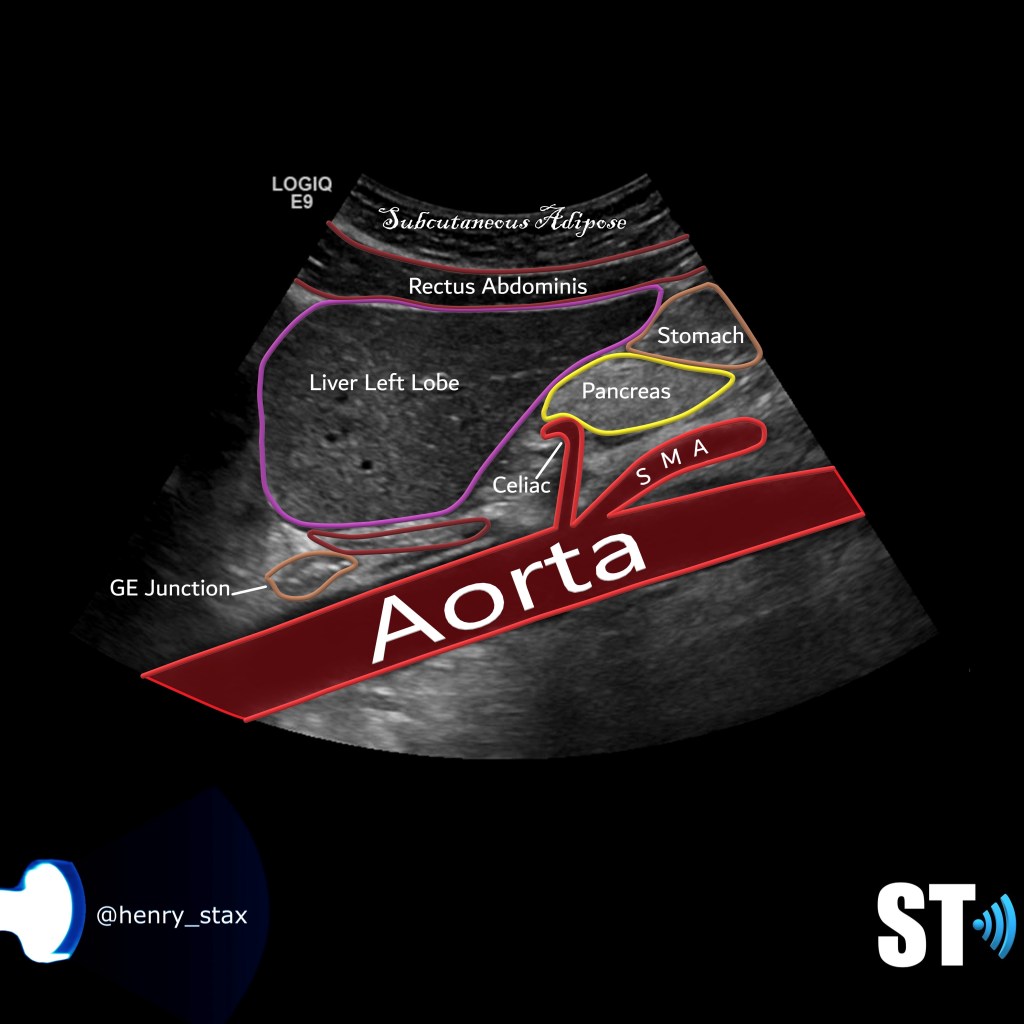
The aorta is the largest artery that carries oxygenated blood from the heart to the viscera and extremities. Here I share with you the protocol for scanning aortic duplex ultrasounds.
Start in the epigastric region at the proximal abdominal aorta and take sagittal grayscale, color Doppler and spectral Doppler and measure the diameter in anterposterior dimensions from outer wall to outer wall, save the images as such.
Sagittal

Transverse
Turn your transducer counterclockwise and take transverse images with and without color.

Middle Aorta
Sagittal
Continue to the mid abdominal aorta and take sagittal grayscale, color Doppler and spectral Doppler and measure the diameter in anterposterior dimensions from outer wall to outer wall, save the images as such.

Transverse
Turn your transducer counter clockwise and take transverse images with and without color Doppler.

Distal Aorta
Sagittal
Continue to the distal aorta and take sagittal grayscale, color Doppler and spectral Doppler and measure the diameter in anterposterior dimensions from outer wall to outer wall, save the images as such.

Transverse
Turn your transducer counter clockwise and take transverse images with and without color Doppler.

Bifurcation
Sagittal
Then move on the the bifurcation, to achieve this image you’ll have to move right later at the umbilical level and angle obliquely towards the vertebrae.

Transverse
Turn the probe counterclockwise and take transverse images with and without color.

Right Common Iliac Artery
Sagittal
Then move on to the right iliac fossa and scan the right iliac artery, take sagittal grayscale, color Doppler and spectral Doppler and measure the diameter in anterposterior dimensions from outer wall to outer wall, save the images as such.

Transverse
Turn the probe counterclockwise and take transverse images with and without color.

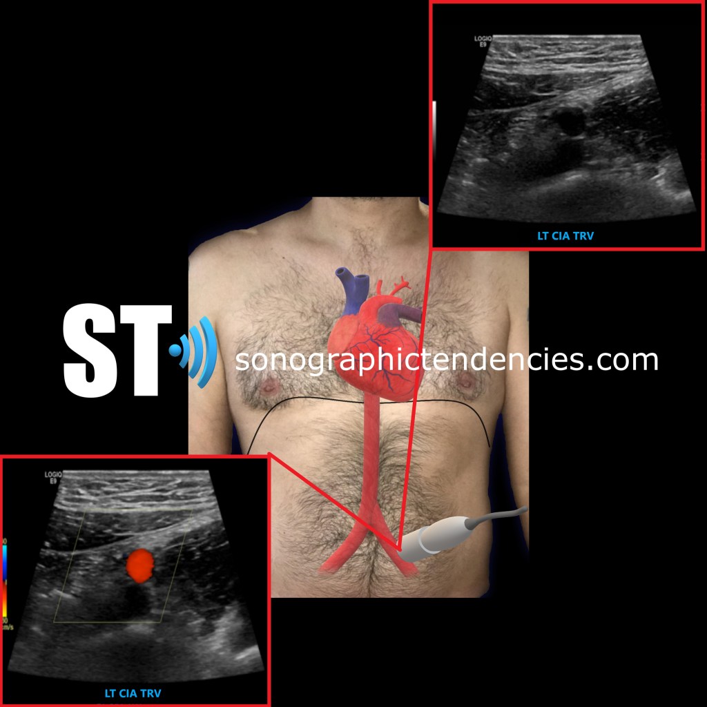
Left Common Iliac Artery
Sagittal
Finally finish your exam by interrogating the left iliac artery. Same scheme as before sagittal, grayscale, color Doppler and spectral Doppler also measure.

Transverse
And transverse, with and without color.

That was so educational Henry, Thank you. I’ve seen some sonographers use sector Array probe (Echo). What is your opinion about using sector array?
Thanks
LikeLike
Using a sector for abdomen is fine especially on bigger patients. The resolution might suffer a little bit but otherwise not a problem
LikeLike