

Here is the protocol for the complete renal ultrasound.
BLADDER
Sagittal
With a full bladder begin at midline and take right and left lateral images in sagittal. Measure the bladder wall per protocols or indication. A normal adult bladder can hold about 470 ml of urine or about 2 cups.

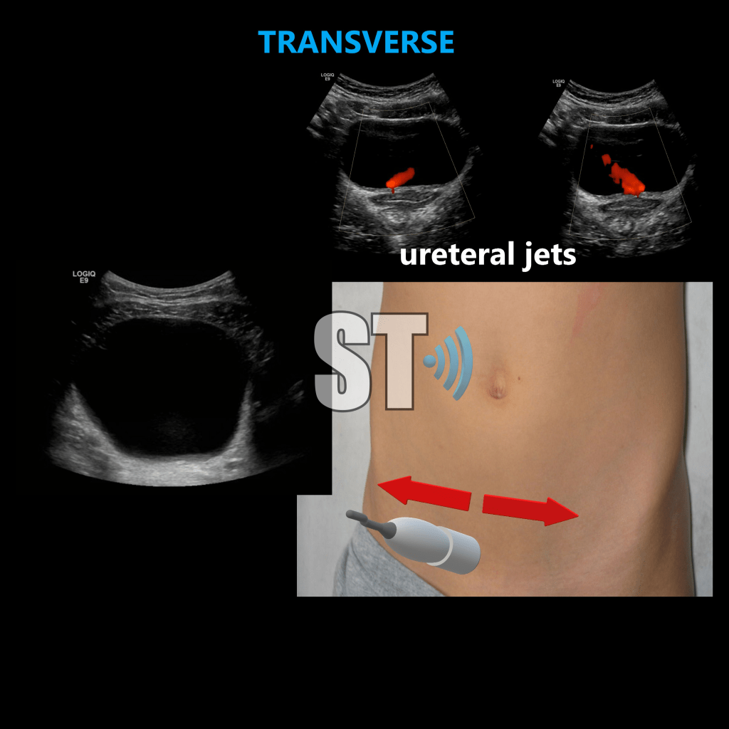
TRANSVERSE
Turning your transducer counter clockwise you’ll be in the transverse plane. You may sweep from superior to inferior and take images accordingly. If clinically warranted watch for and take ureteral jets by placing doppler over the ureteral orifice (the ureteral jets are the periodic efflux of urine into the bladder by ureteral peristalsis).
RIGHT KIDNEY
Sagittal
Using a curved or sector transducer take sagittal images using the liver as an acoustic window. Aske the patient to breath deeply and hold it to improve visualization. Compare the echogenicity of the kidney to the liver, the liver should be more echogenic.Take medial, lateral and midline sagittal images, color doppler is not required by every laboratory, use if clinical indicated or warranted, (i.e. trying to rule out pyelonephritis, mild hydronephrosis versus renal vein at the hilum, making a calculus exhibit the twinkle artifact etc…)

TRANSVERSE
In transverse take superior, mid and inferior pole images as shown in the image below. Again use patient breathing to assist in visualization.

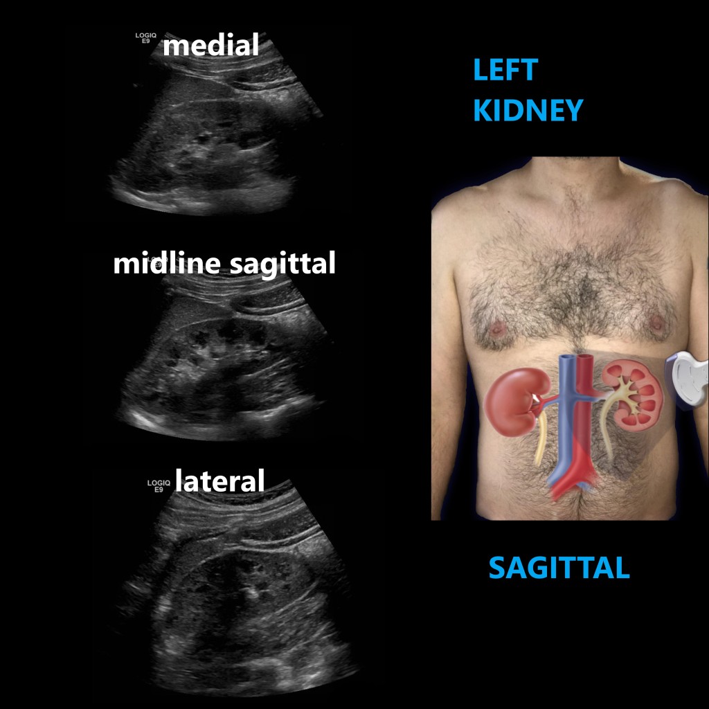
LEFT KIDNEY
Sagittal
Repeat the steps for the right kidney. The spleen not being as large as the liver offers a poor window for the left kidney, which is also close to the stomach. As such visualization can be more difficult of the left kidney. Having the patient holding their inspiration can help as can using a sector probe and scanning through the intercostal spaces. Another technique is asking the patient to turn right lateral oblique.
Transverse
As on the right so goes the left, take superior, mid and inferior pole shots of the left kidney, compare the echogenicity of the left kidney to the spleen. The spleen should be more echogenic than the kidney.

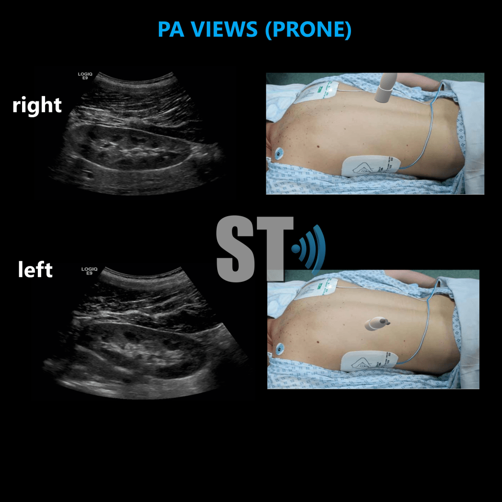
PRONE POSTEROANTERIOR (PA) VIEWS
Lastly have the patient lay on their abdomen, the prone position, and scan the kidneys from the back. They will be located just lateral to the spine on either side. These views are not required in every laboratory, however they can assist by offering alternative views and opportunities to discover pathology that may go missing in traditional anterior views. Take transvers and sagittal views. In slimmer patients use a linear probe and you’ll be pleased at great images you can acquire!

[…] Renal Ultrasound Complete Protocol […]
LikeLiked by 1 person
Extremadamente practico y útil para los médicos que se inician en la ecografia clínica.
LikeLiked by 1 person
Gracias por el comento!!!
LikeLiked by 1 person
Nice video. As a mid pole is a non-excisting place in real world, mid, or inter polar could be a better term. The presence of a jet does not rule out uretral or kidney stones.
LikeLike
Haha yes to the interpolar, and very true about presence of jets not excluding calculi.
LikeLike