

Protocol image sequence (Right side only repeat images on left testis for complete protocol).


Transverse
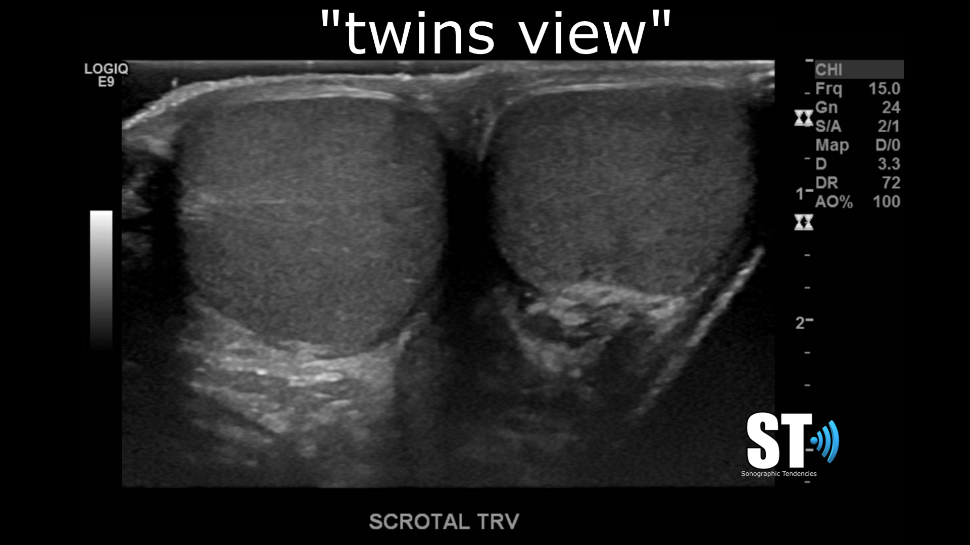
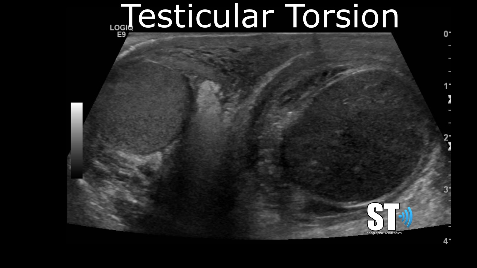
Begin in transverse what’s know colloquially as the “twins” view.
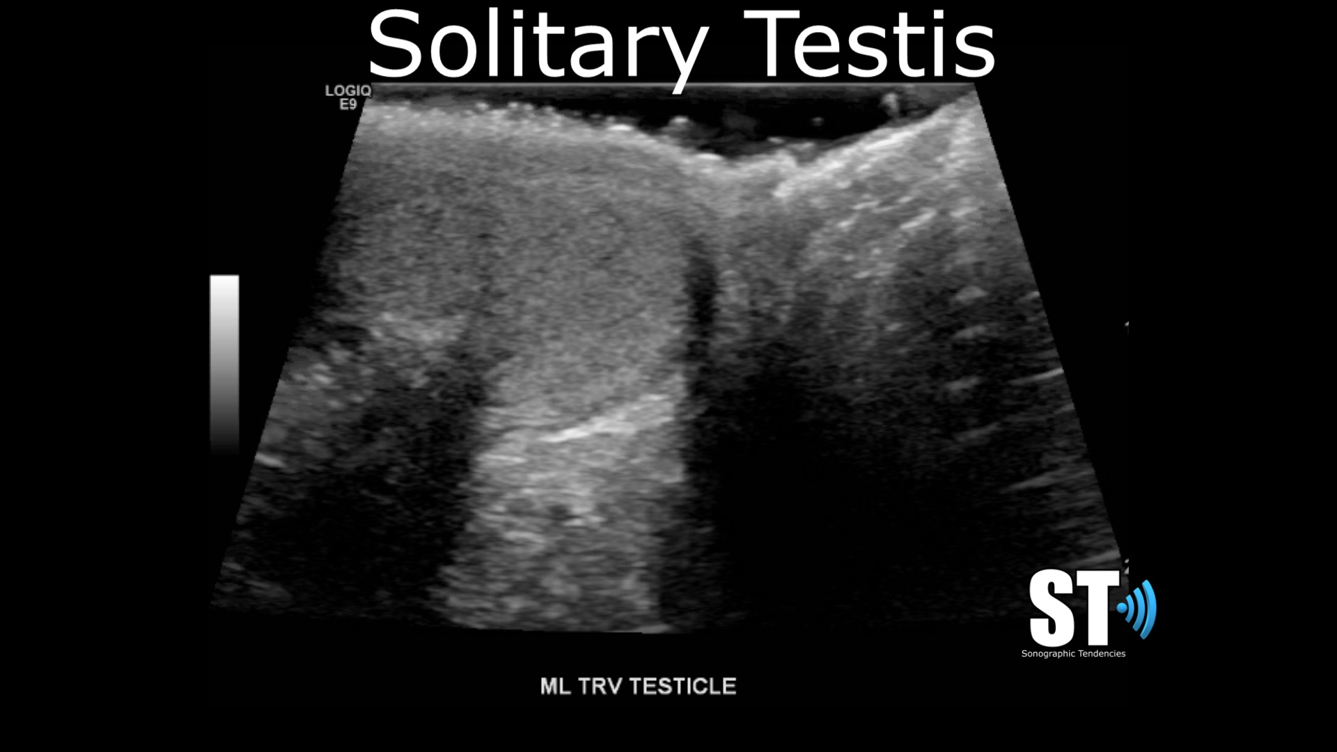
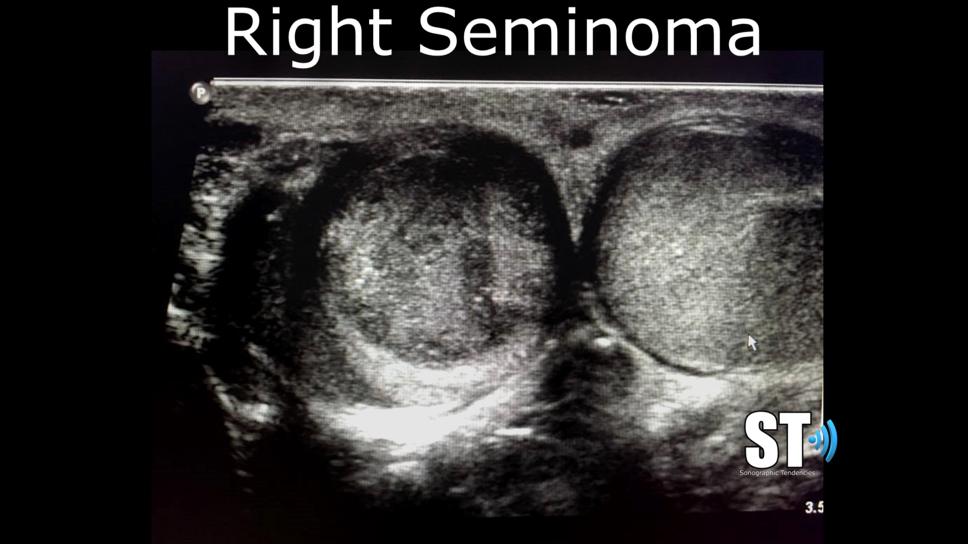
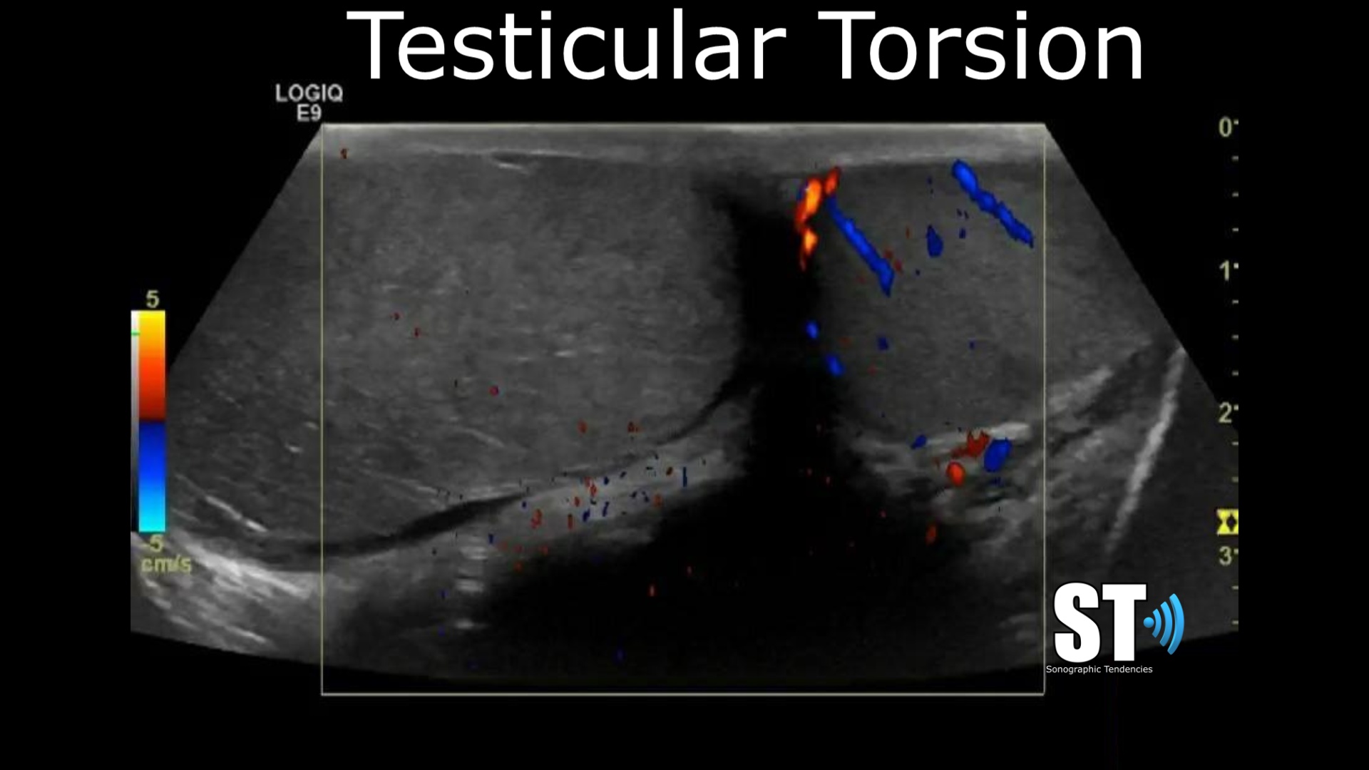
This view is great for determining quickly symmetry, position and echotexture. With color doppler you can determine whether there is testicular torsion. In cases of undescended testis you can show a solitary or even empty scrotum if the cryptorchidism is bilateral. Also if there is any testicular lesions they may be apparent form your first image.
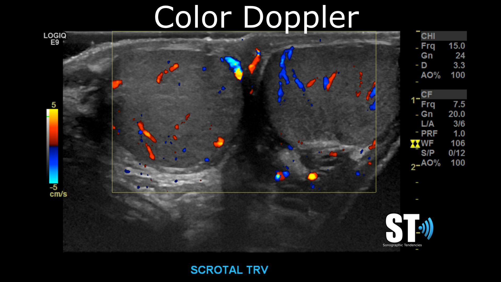
Color Doppler
In the same transverse view use color doppler to confirm to see if the blood flow is symmetric. In cases of epididymo-orchitis there will be hyperemia, or increased blood flow to the affected testis. Again if there is testicular torsion it will be apparent.
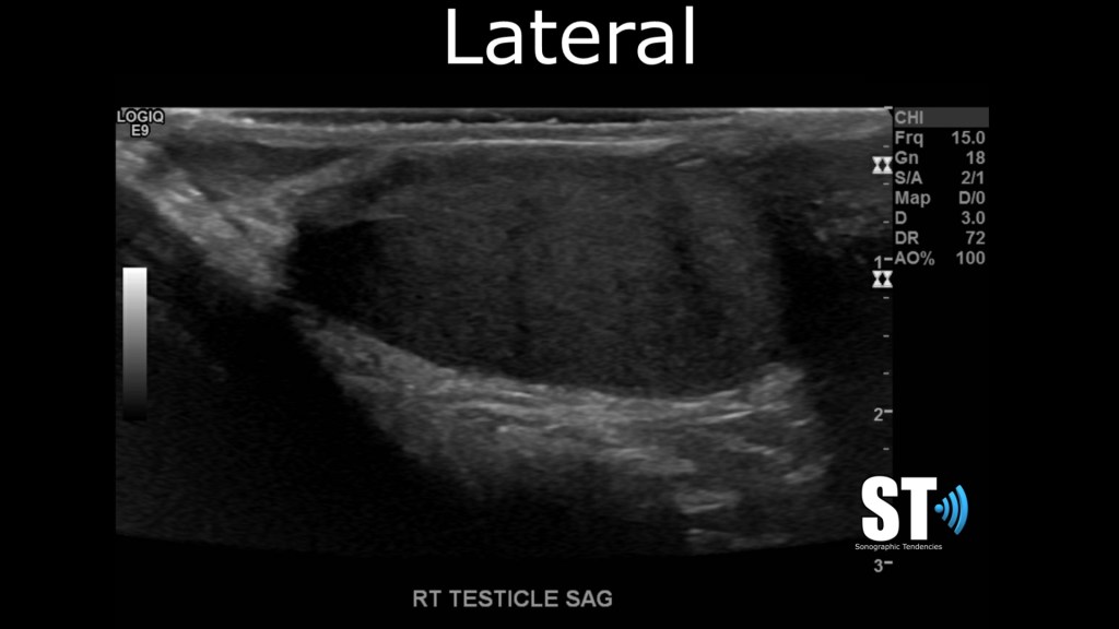
Sagittal
Once your done with your initial transverse images begin your sagittal images with the right testicle. Capture an image in sagittal midline, sagittal lateral and medial. Show the mediastinum testis in the medial view. Assess for homogenous echotexture, hydroceles, tumors etc..


Sagittal Lateral and Medial


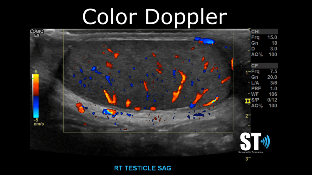
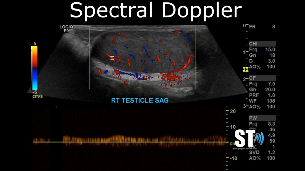
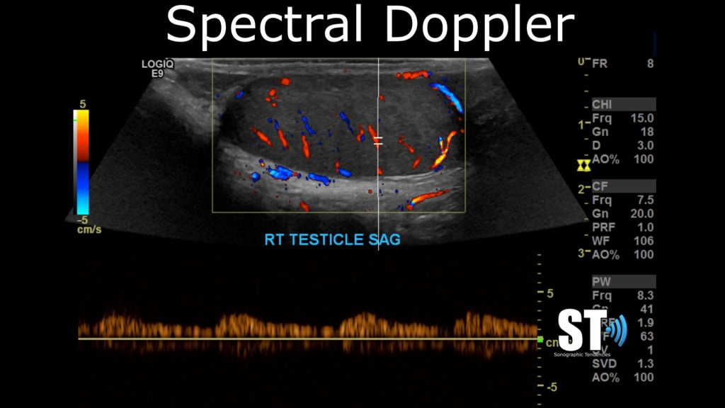
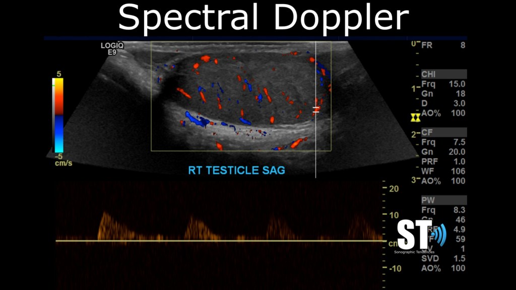
Color and Spectral Doppler
After you’re done with you sagittal grey scale images, use color and spectral doppler to interrogate the testes and take spectral tracings at the superior, mid and inferior sections of the testicle.
Measure
Next take measurements of length x width x height.

Normal adult testes measure approximately 3-5 cm (length) x 2-4 cm (width) x 3 cm (height) and normal range of volume is 12.5-19 mL 2.
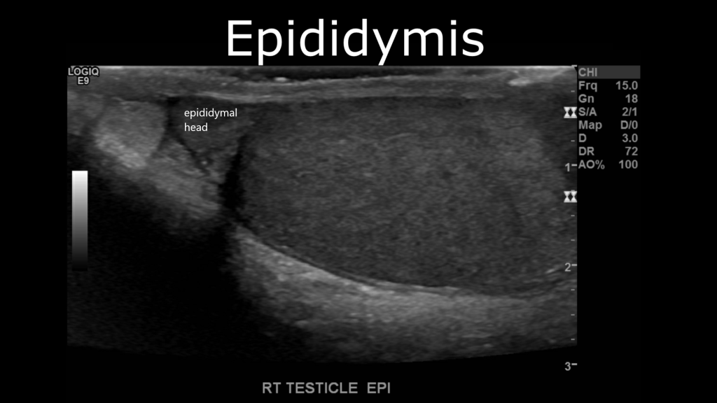
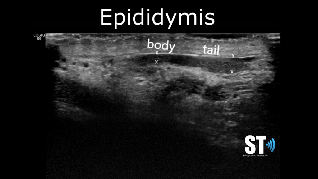
Epididymis
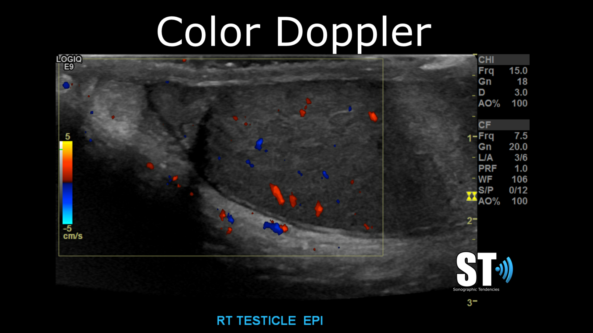
In sagittal image the epididymis, check for homogeneity, and look out for epididymal cysts and spermatoceles. Depending on your institution you may or may not have to measure the dimensions of the epididymis. Take images with color doppler. In cases of epididymitis there will be enlargement, hyperemia and fat edema.


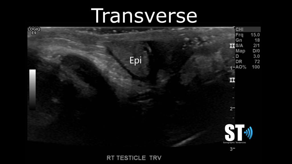
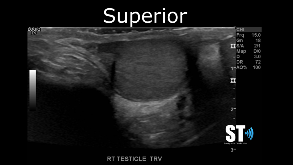
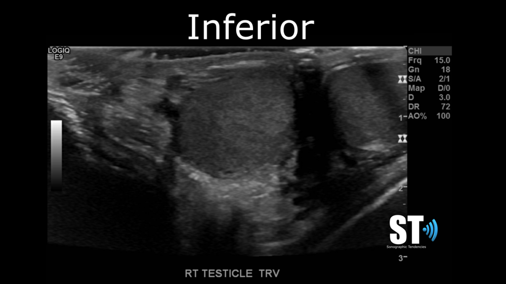
Transverse
In transvers beginning at the superior pole take images of starting at the epididymal head and capture images of the superior, mid section and inferior poles of the testis.


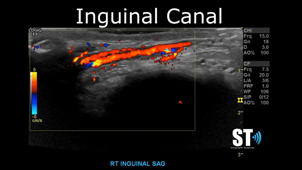
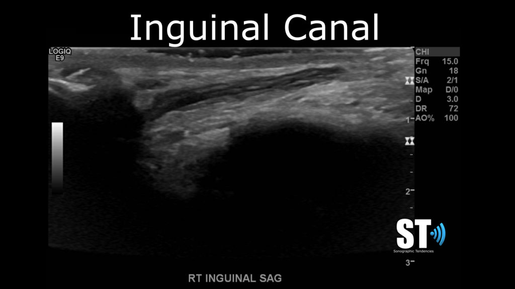
Inguinal Canal
When you’ve completed imaging the testicle, begin scanning the inguinal canals and inspect the spermatic cord and pampiniform plexus to detect varicoceles or inflammatory conditions like funniculitis/vasitits. Also here is an excellent place to evaluate for hernias that are present and haven’t reached the scrotum. Valsalva maneuver can assist in clinching the diagnosis of inguinal hernia or varicocele.

Valsalva
Here’s a clip of a varicocele. You can see the increase in blow flow after Valsalva maneuver.


[…] Testicular/Scrotal Doppler Protocol […]
LikeLike
What is the correct way to measure the epididymis?
LikeLike
When you do the left side – is the epididymus head lateral or medial (will it be on the right or left side of screen)? I have seen it done both ways I thought and now I am very confused. I thought the head was always lateral and superior and the tail was always lateral inferior but yesterday I saw someone look at the left epididymus medially…. I need to do this exam this week alone – help me! thanks!!!!!!! I love you and your videos and your instagram and am a huge fan and think you are hilarious and so smart btw. ❤
LikeLike