

The thyroid gland is an endocrine gland in the neck consisting of two lobes connected by the isthmus. The thyroid is located at the front of the neck, just below the laryngeal prominence of the thyroid cartilage.

Begin in the midline neck transverse below the thyroid cartilage.


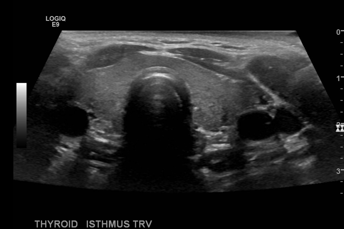
Take image of the thyroid at midline, sweep superior and inferior and measure the isthmus in anteroposterior dimension. Also place color doppler over the gland.


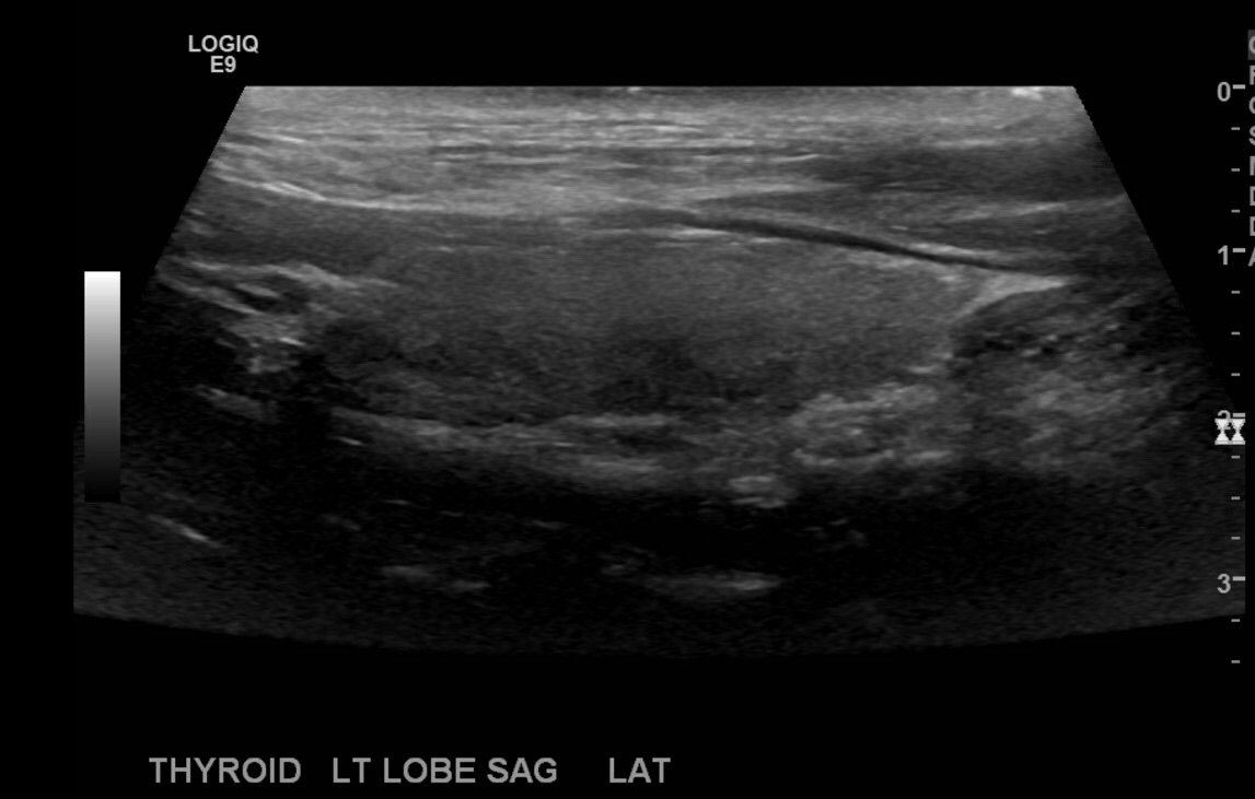
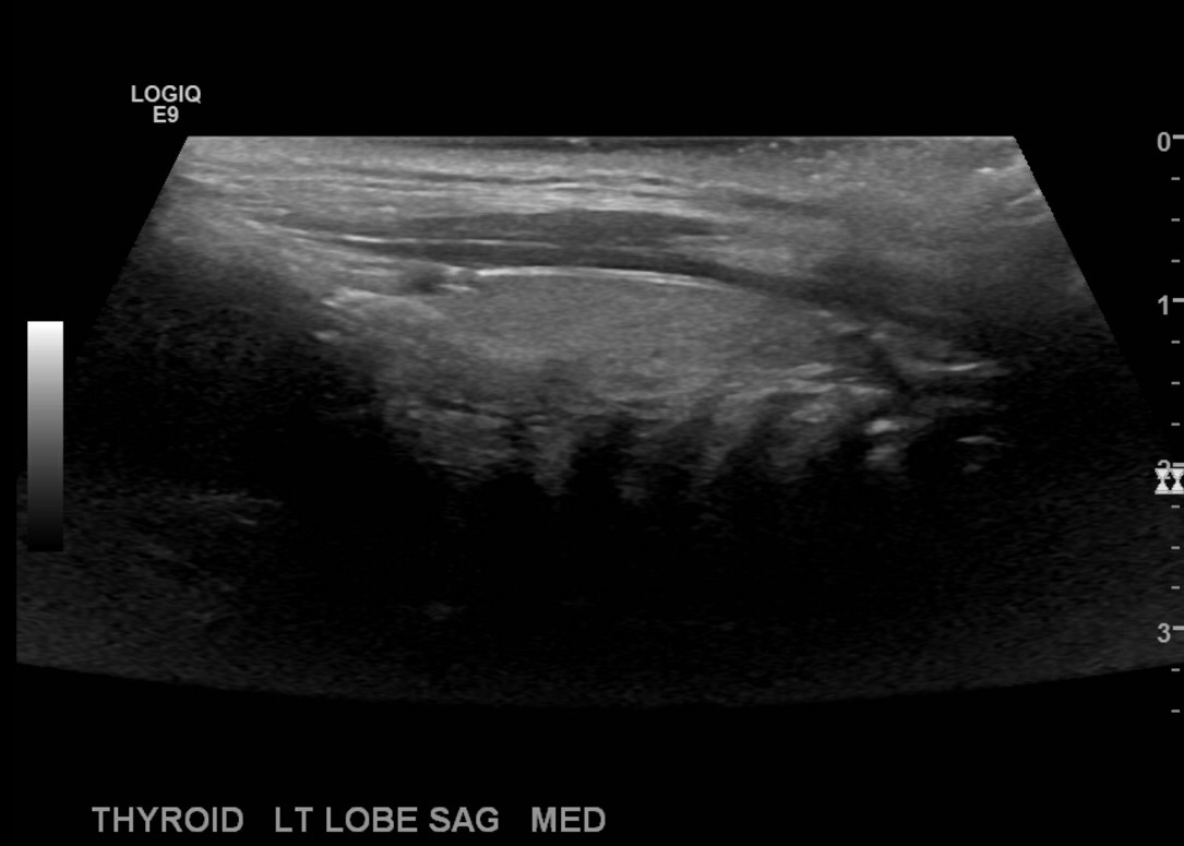
Then go to the right neck and take sagittal images in middle of the right thyroid lobe, lateral and medial.


Then measure the thyroid length x width x height. Normal thyroid lobe dimensions change from birth into adulthood: the length (L or craniocaudal) diameter is 1.8 to 2.0 cm in newborns and 4.0 to 6.0 cm in adults, while the A-P dimension measures 0.8 to 0.9 cm in newborns and 1.3 to 1.8 cm in adults.

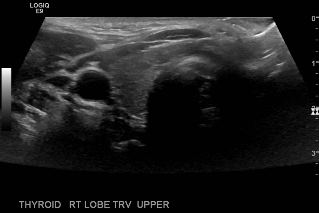
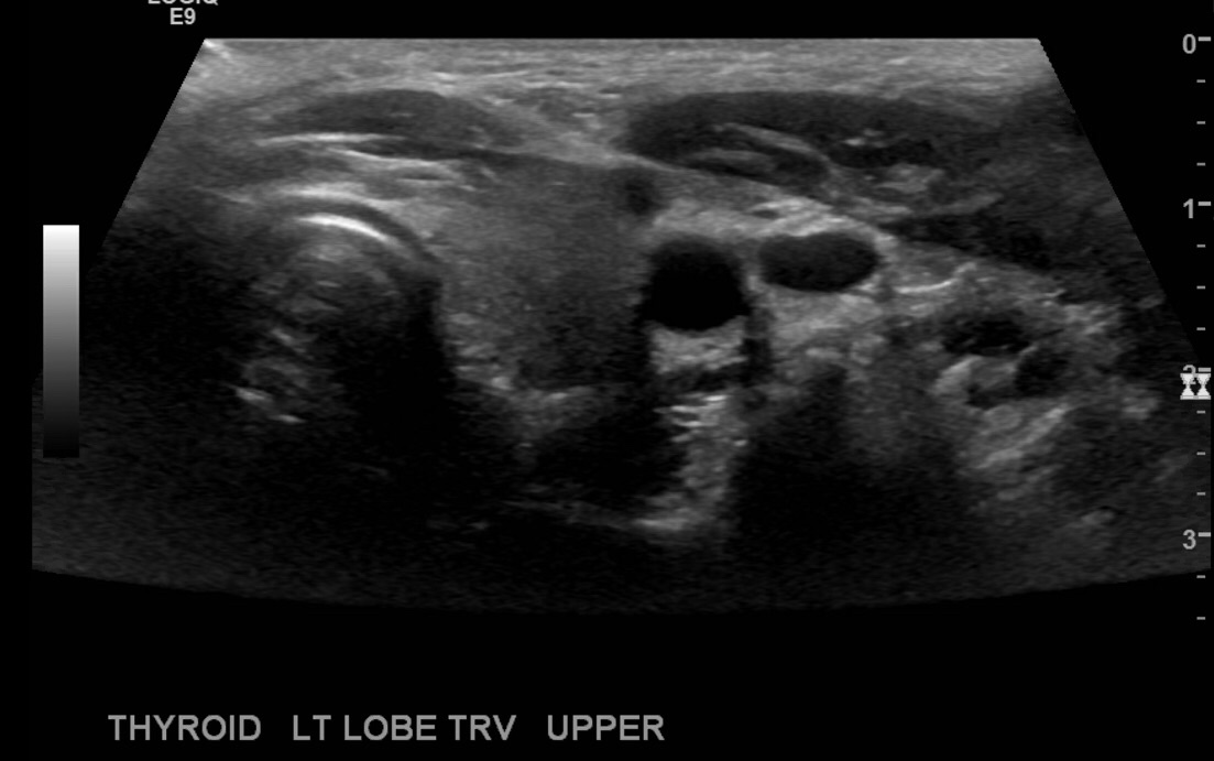
Turn the transducer counterclockwise and take transverse images at the middle of the rt lobe, superior and inferior.


Repeat the process on the left.
Go to the right neck and take sagittal images in middle of the right thyroid lobe, lateral and medial.

Then measure the left thyroid lobe, length x width x height.


Turn the transducer counterclockwise and take transverse images at the middle of the rt lobe, superior and inferior.

Document abnormalities as you encounter them, with measurements, color and spectral Doppler as necessary.

Amazing . Crystal clear explaination.
LikeLike
Thank you!!
LikeLike
Thank you so much for this. Midterms week!!!
LikeLike