


Umbilical Artery Doppler Study
Umbilical artery Dopplers are monitored for a variety of reasons in MFM. This is typically a part of a fetal well being assessment. We use the umbilical artery Doppler in the 2nd and 3rd trimester for growth restriction which helps us to determine placental insufficiency. According to Radiopeadia, “Umbilical artery Doppler assessment has been shown to reduce perinatal mortality and morbidity in high-risk obstetric situations.” We also use it in MFM to monitor Monochorionic-Diamniotic twins for twin-to-twin transfusion syndrome (TTTS).
The umbilical cord itself is made of 2 umbilical arteries (mean width of 2.4mm) and one umbilical vein (neam width of 8mm). The umbilical cord is ensheathed in Wharton’s jelly for protection.
For assessment of the umbilical cord we try and sample the umbilical cord to as close as you can get to a 0º angle. If you sample the umbilical cord too close to the placental cord insertion (PCI) or to the abdominal cord insertion (ACI) your numbers can be abnormally elevated. My practice prefers we measure a free loop of floating cord, but I’ve known other offices that want measurements at the PCI, free floating, and at the ACI. Your practice’s protocols will dictate where you should sample from.
The flow in the umbilical cord should always have a “forward” flow. Forward flow means flow that is towards the PLACENTA. For those of you that have taken your fetal echo boards, this was highlighted for us. We learned that the Umbilical artery goes TOWARDS the Placenta and the Umbilical Vein goes TOWARDS the Baby!
UMBILICAL ARTERY ——> PLACENTA
UMBILICAL VEIN ——> BABY
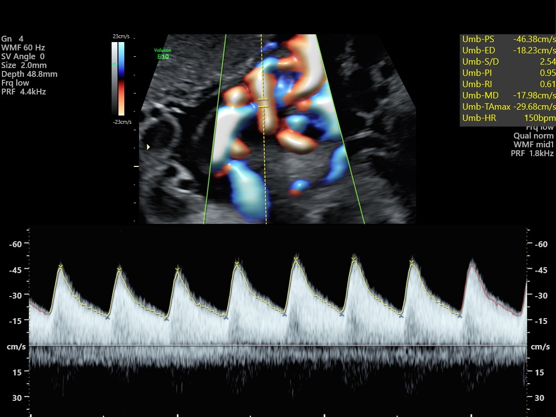
The S/D ratio is higher early in pregnancy and decreases as the baby grows. You can sometimes see absent end diastolic flow in fetuses less than 15 weeks, which can be normal.
According to Radiopeadia:
- At 20 weeks – 50% for S/D ratio is 4
- At 30 weeks – 50% for S/D ratio is 2.83
- At 40 weeks – 50% for S/D radio is 2.18
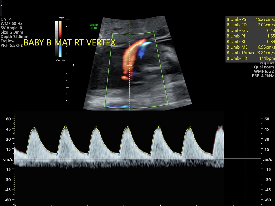
In growth-restricted fetuses and fetuses developing intrauterine distress, the umbilical artery blood velocity waveform usually changes in a progressive manner as below:
- reduction in end-diastolic flow: increasing RI values, PI values, and S/D ratio
- absent end-diastolic flow (AEDF): RI = 1
- reversal of end-diastolic flow (REDF)
Normal Umbilical artery Doppler- “Sawtooth pattern”
An abnormal umbilical artery Doppler can have a high S/D ratio.
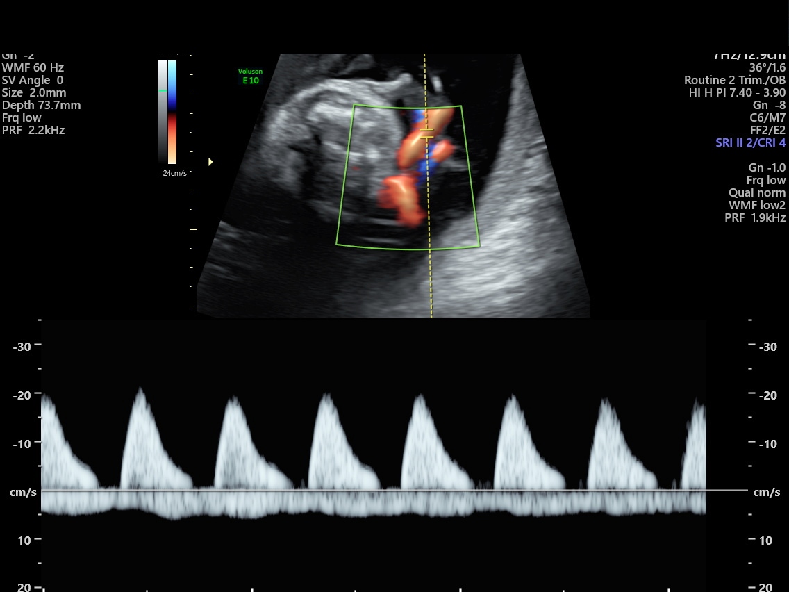
An abnormal Umbilical artery can have absent end diastolic flow (AEDF). AEDF in mid to late pregnancy usually occurs as a result of placental insufficiency.
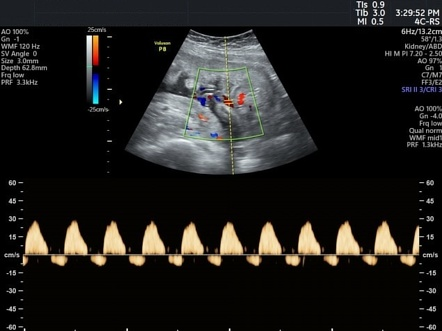
Or an abnormal umbilical artery Doppler can have reversal of the flow. Reversal of fluid is a result of significant increase in resistance to blood flow within the placenta. It is usually related to much larger underlying pathology.
The umbilical artery Doppler can look odd when the baby is breathing.
Author: Marybeth Tomory RDMS (ABD, OB, FE)
Illustrations and Editing Henry Suarez RDMS (ABD, OB), RVT

Marybeth’s Instagram: https://www.instagram.com/sono.eyes/
You can find a helpful calculator online at:
https://www.perinatology.com/calculators/umbilicalartery.htm
Berman, Mimi C., Cohen, Harris L. (1997) Diagnostic Medical Sonography: Obstetrics and Gynecology (v.1) Lippincott Williams & Wilkins (January 1, 1900)
https://radiopaedia.org/articles/umbilical-arterial-doppler-assessment?lang=us
https://radiopaedia.org/articles/absent-umbilical-arterial-end-diastolic-flow-2?lang=us
Image of REDF taken from:
https://radiopaedia.org/articles/reversal-of-umbilical-arterial-end-diastolic-flow
