

Duplex sonography of the extremities is a non-invasive and valuable tool in detecting diseases in the blood vessels of the arms and legs. This post will cover the basic evaluation of the upper and lower extremity arterial systems.
Indications
1. DETECTION OF STENOSIS OR OCCLUSIONS IN SEGMENTS OF THE PERIPHERAL ARTERIES.
2. MONITORING OF SITES OF PREVIOUS SURGICAL INTERVENTIONS, INCLUDING SITES OF PREVIOUS BYPASS.
3. MONITORING OF SITES OF VARIOUS PERCUTANEOUS INTERVENTIONS.
4. FOLLOW-UP OF PREVIOUSLY DIAGNOSED DISEASE.
5. EVALUATION OF ARTERIAL INTEGRITY IN THE SETTING OF TRAUMA.
Anatomy
Having a well rounded working knowledge of the arterial anatomy helps immensely when performing these exams. Let’s begin with the upper system.
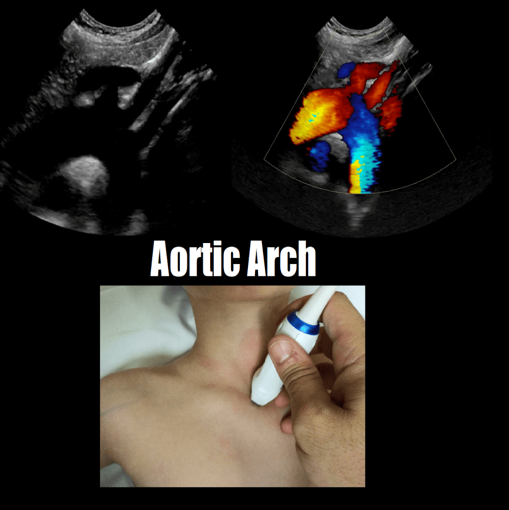
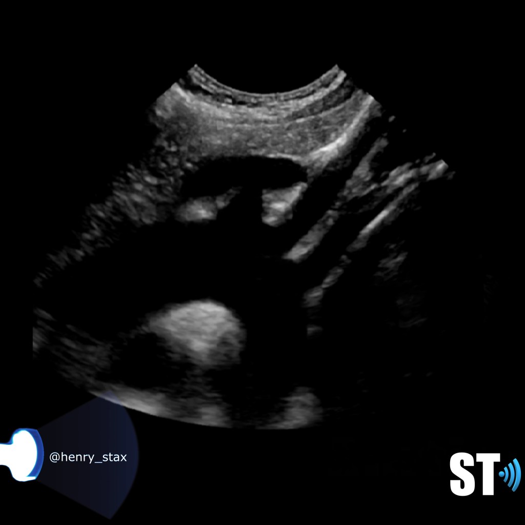
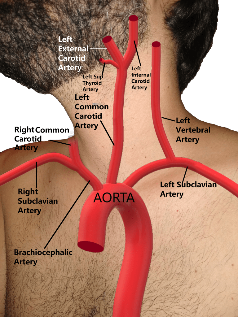
Aorta and its branches
The aorta is the main artery of the body arising from the outflow tract of the left ventricle. It is shaped like an umbrella handle or a candy cane. The first portion is the ascending aorta which then curves to become the aortic arch. Here there are three consecutive vessels that arise and course cephalad. The first is the brachiocephalic or innonimate artery which further bifurcates into right subclavian and common carotid arteries. Next is the left common carotid artery followed immediately by the left subclavian artery.

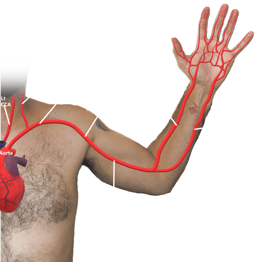
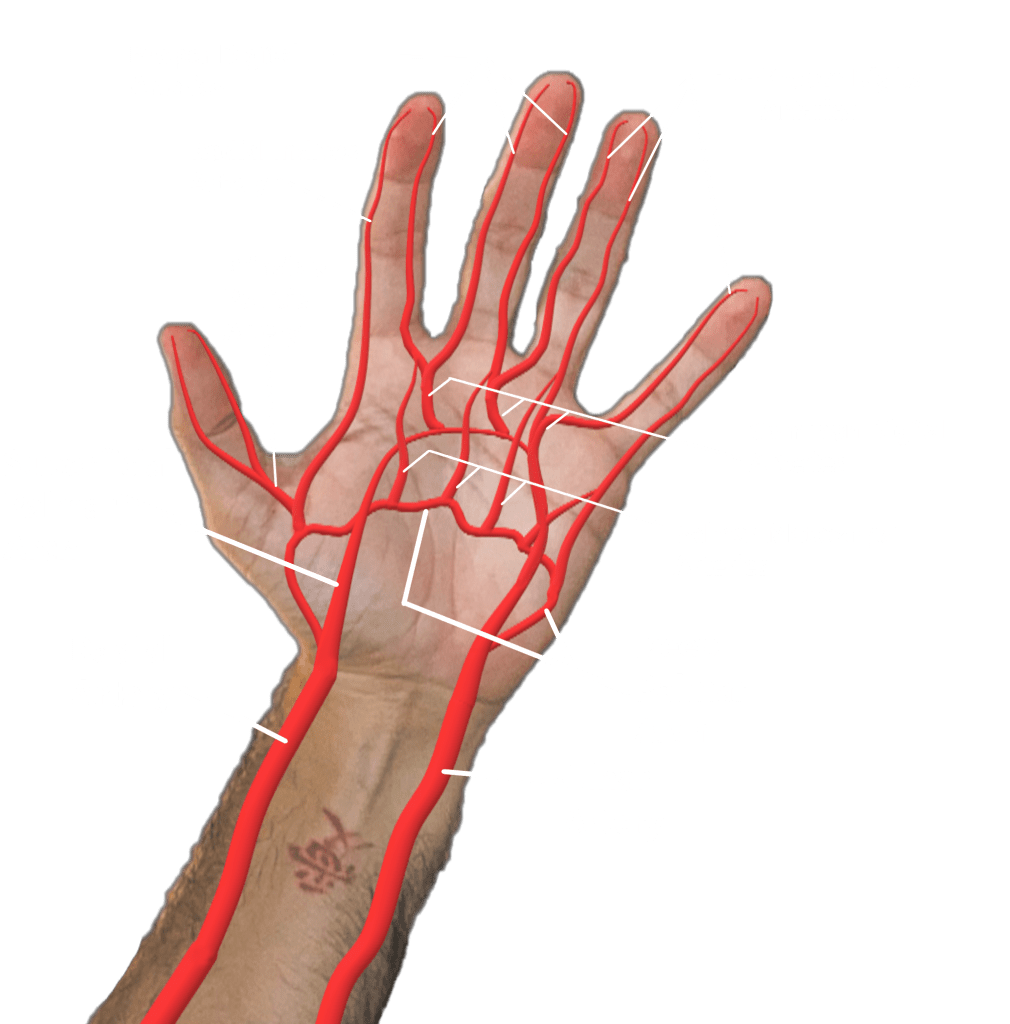
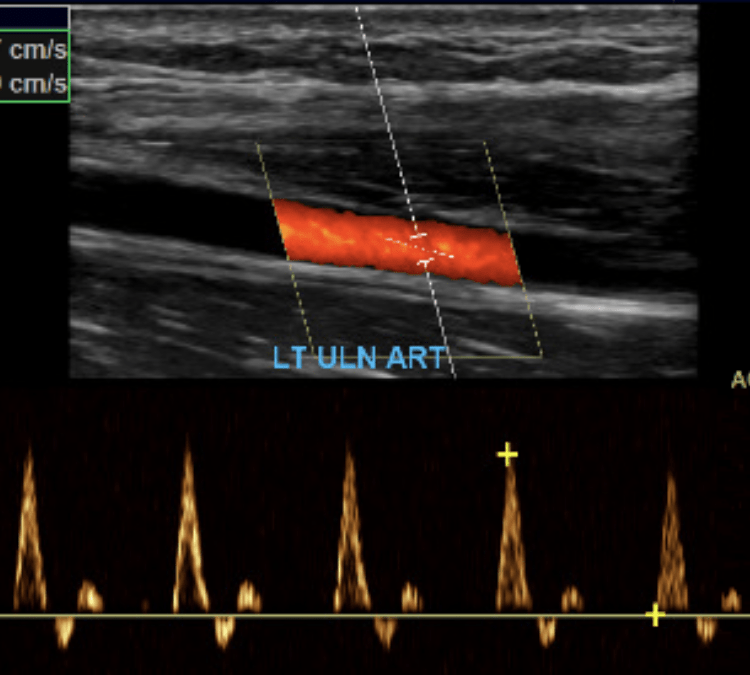
Upper Extremity
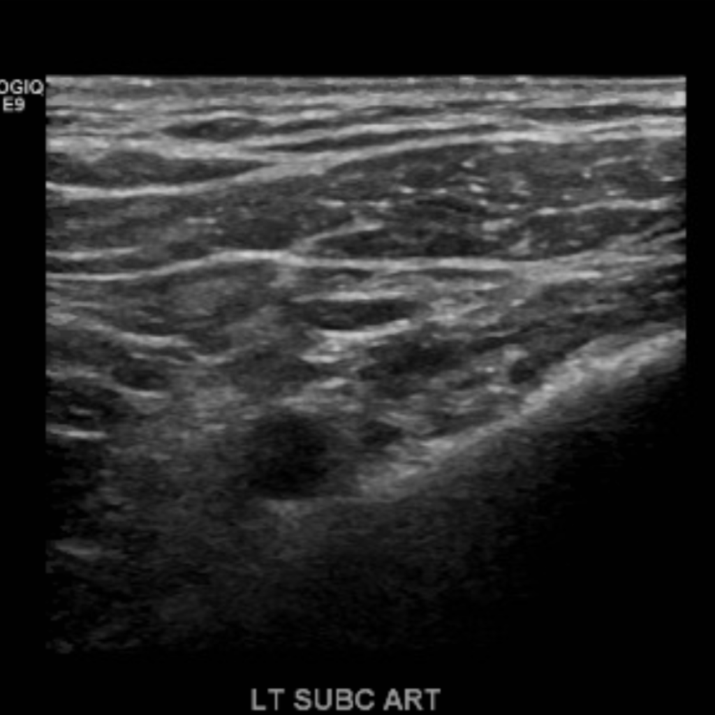
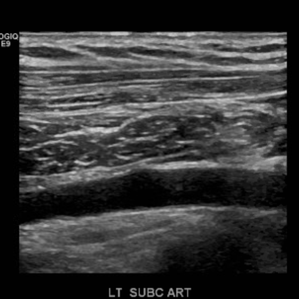
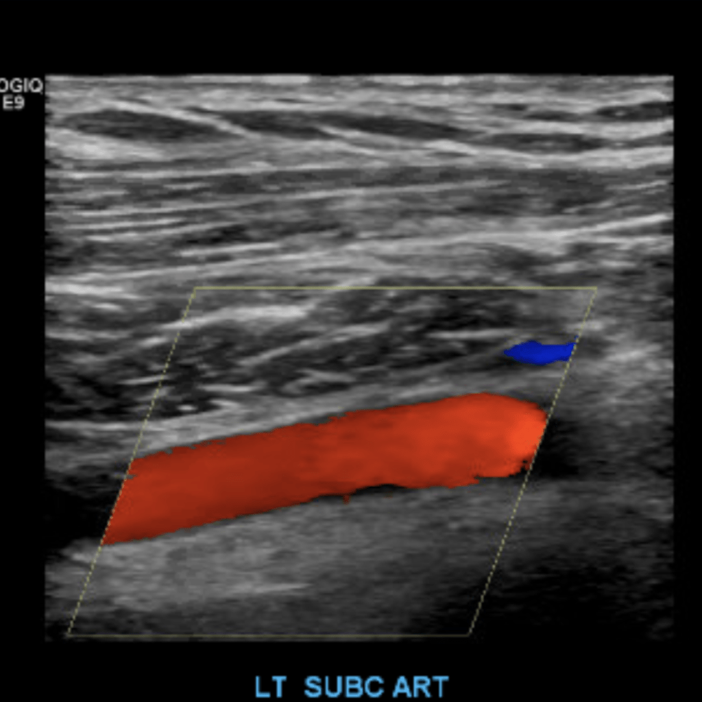
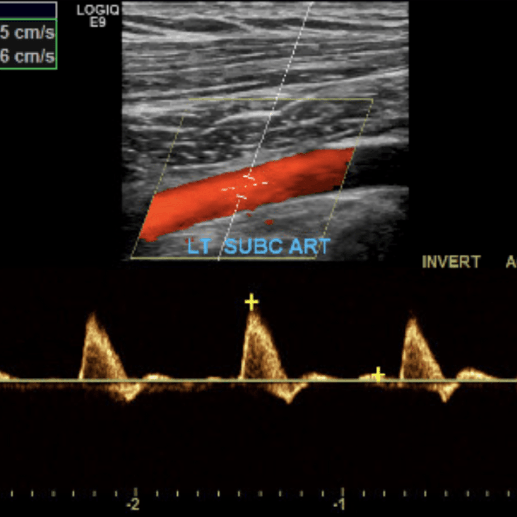
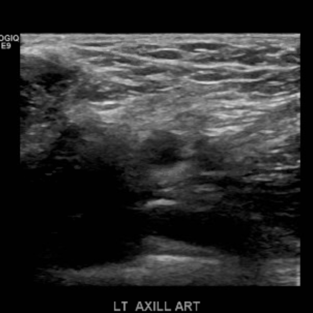
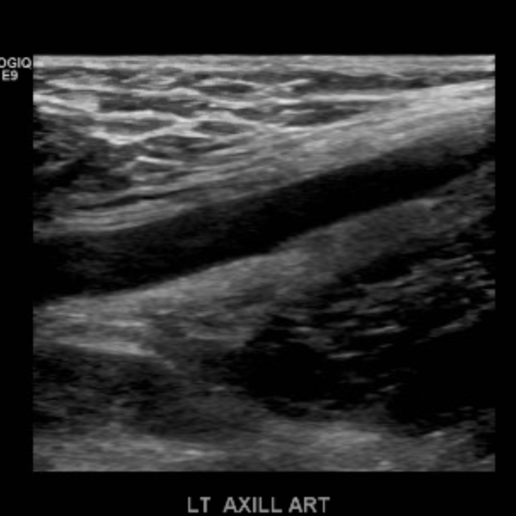
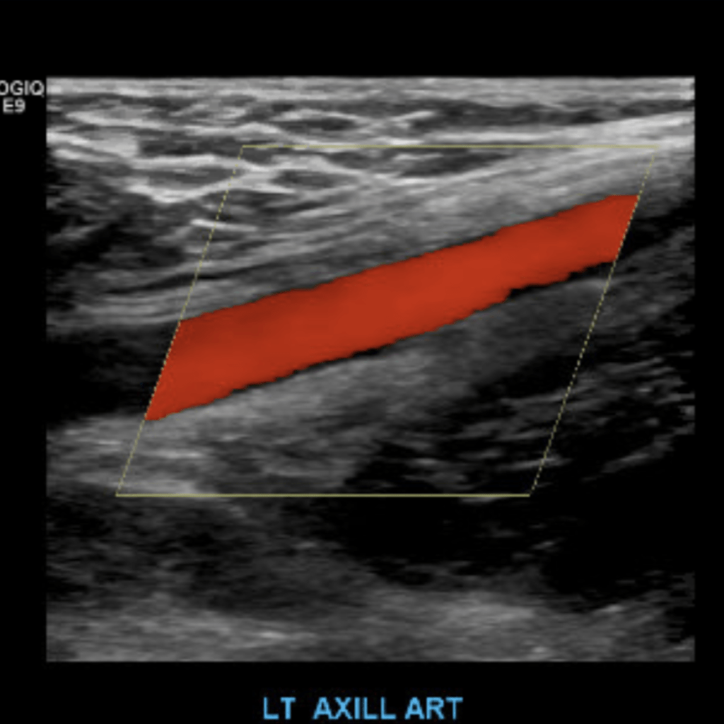
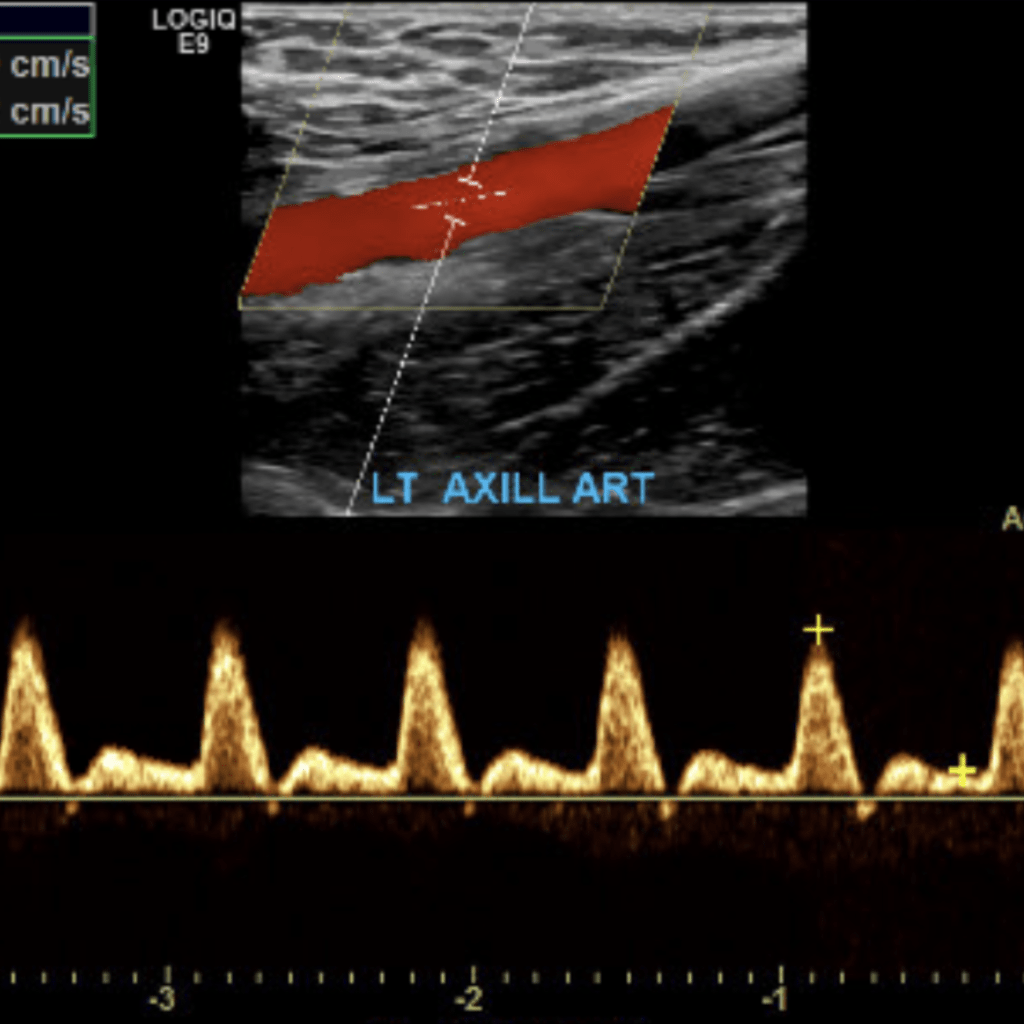
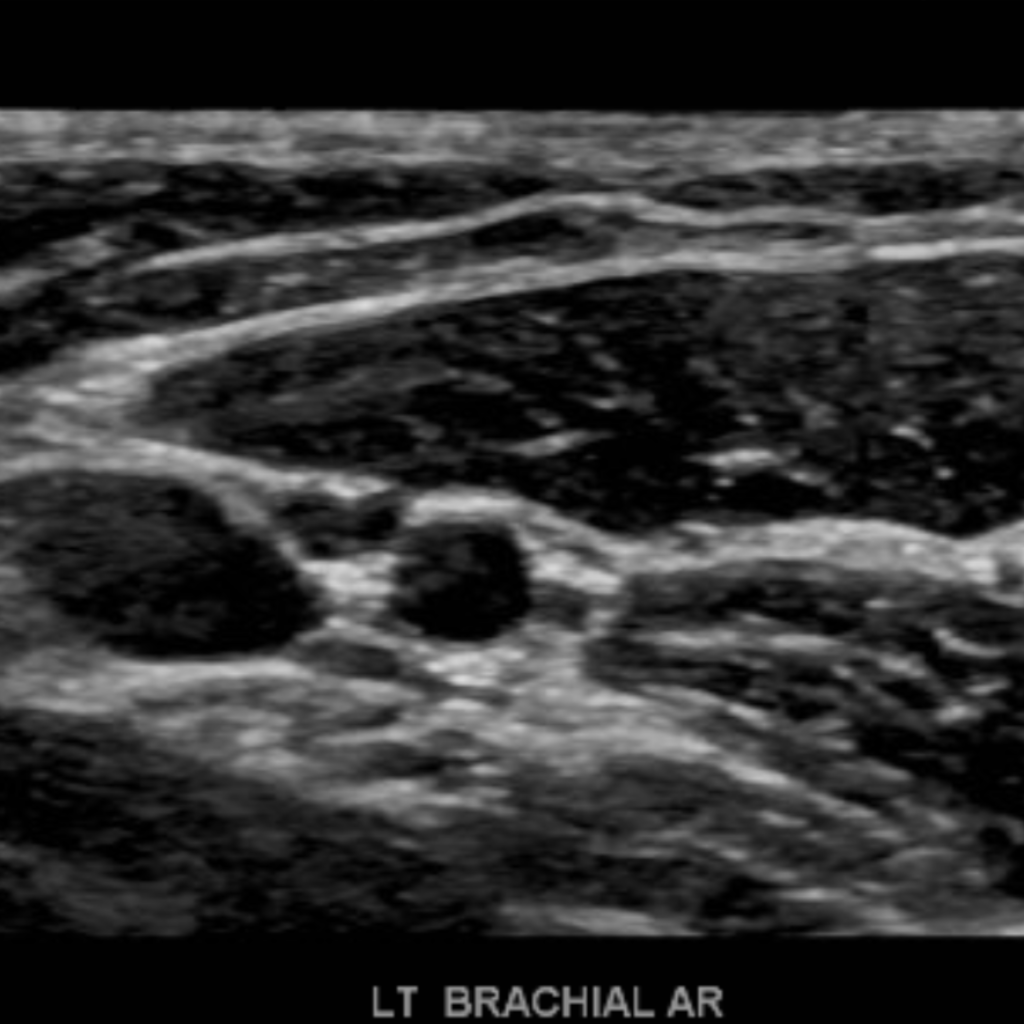
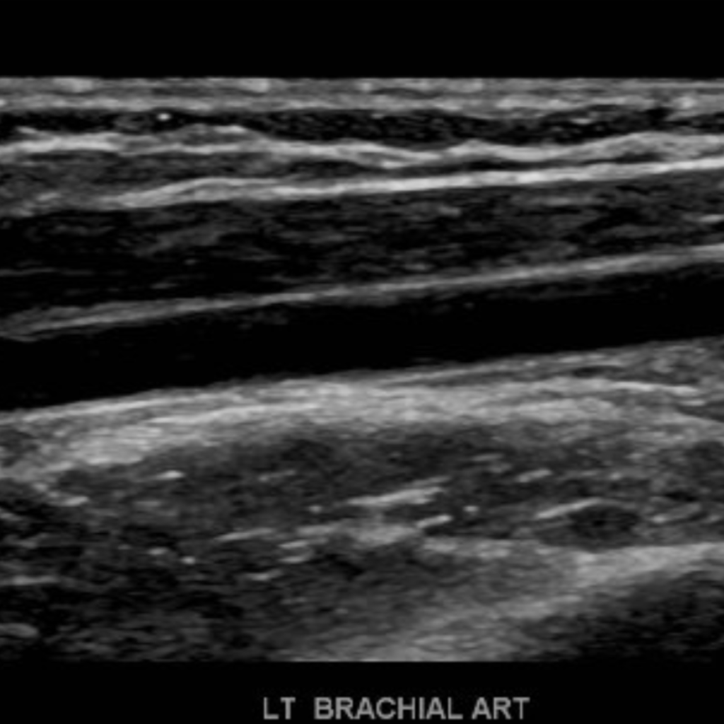
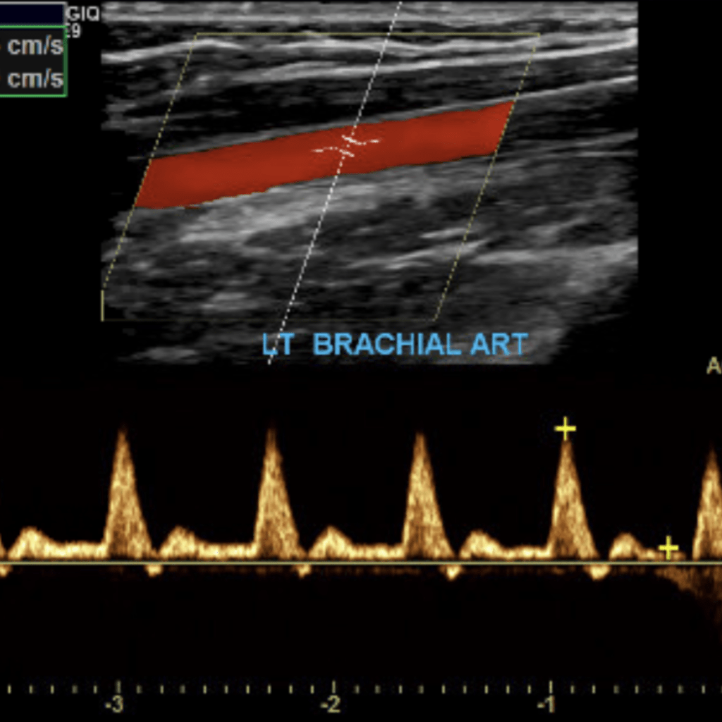
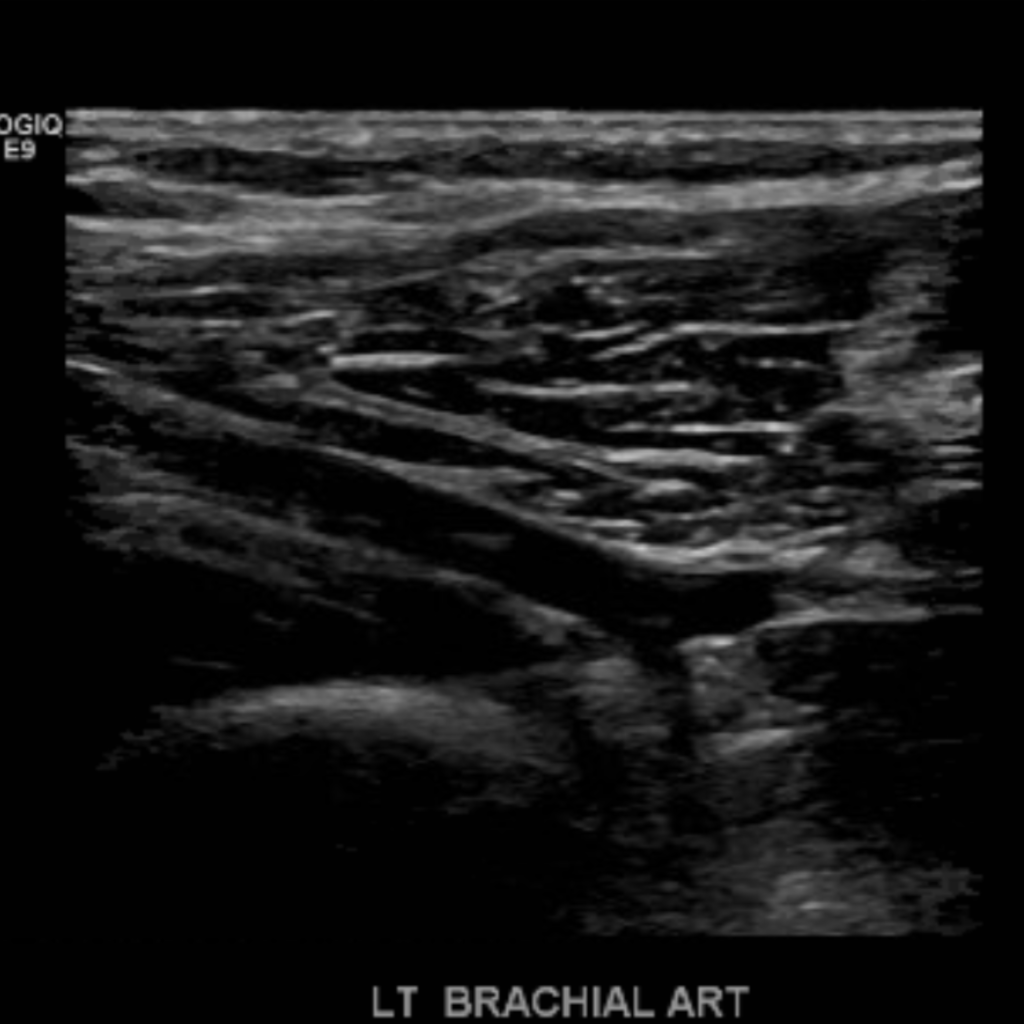
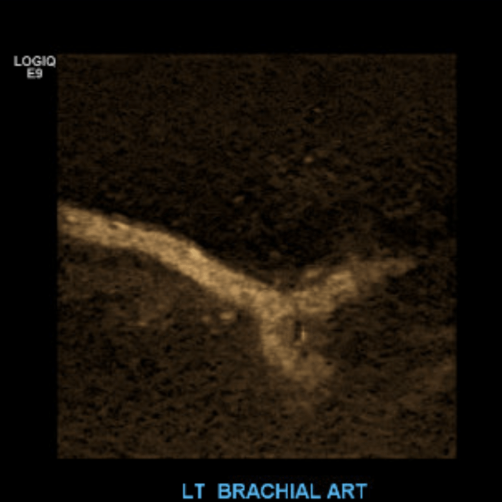
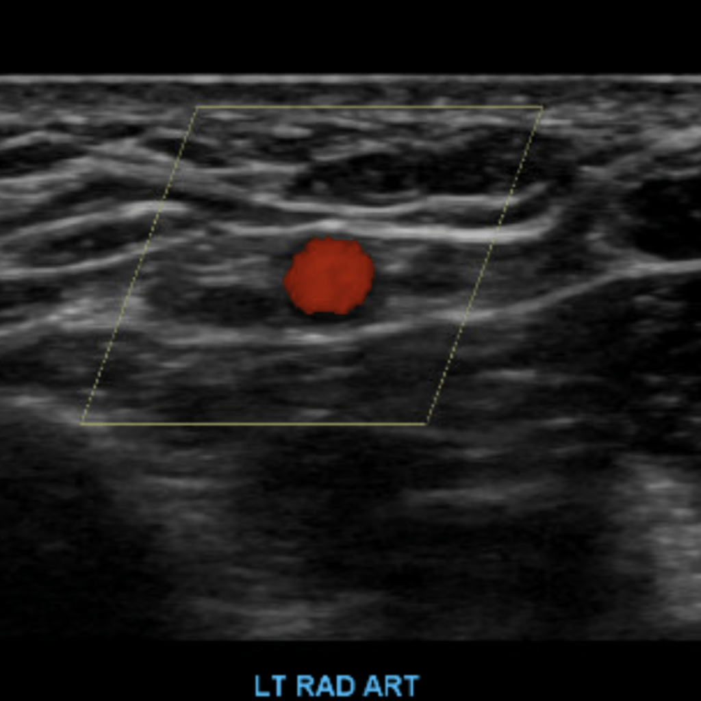
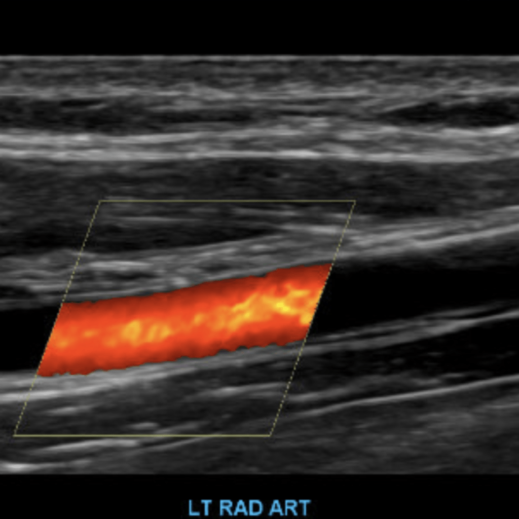
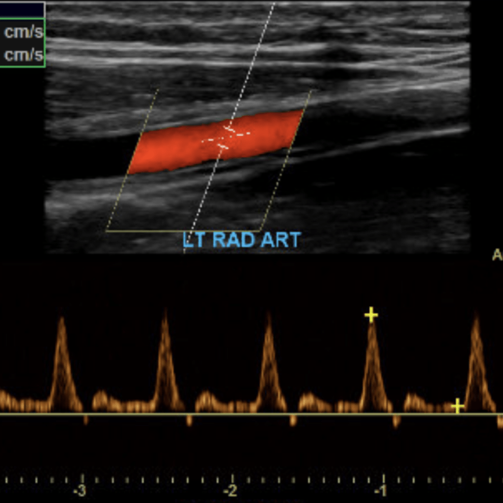
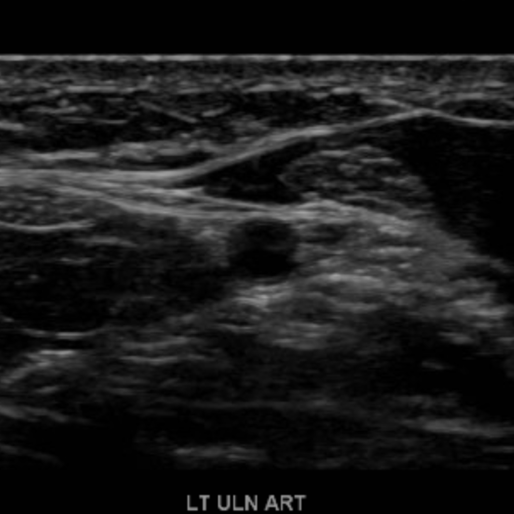
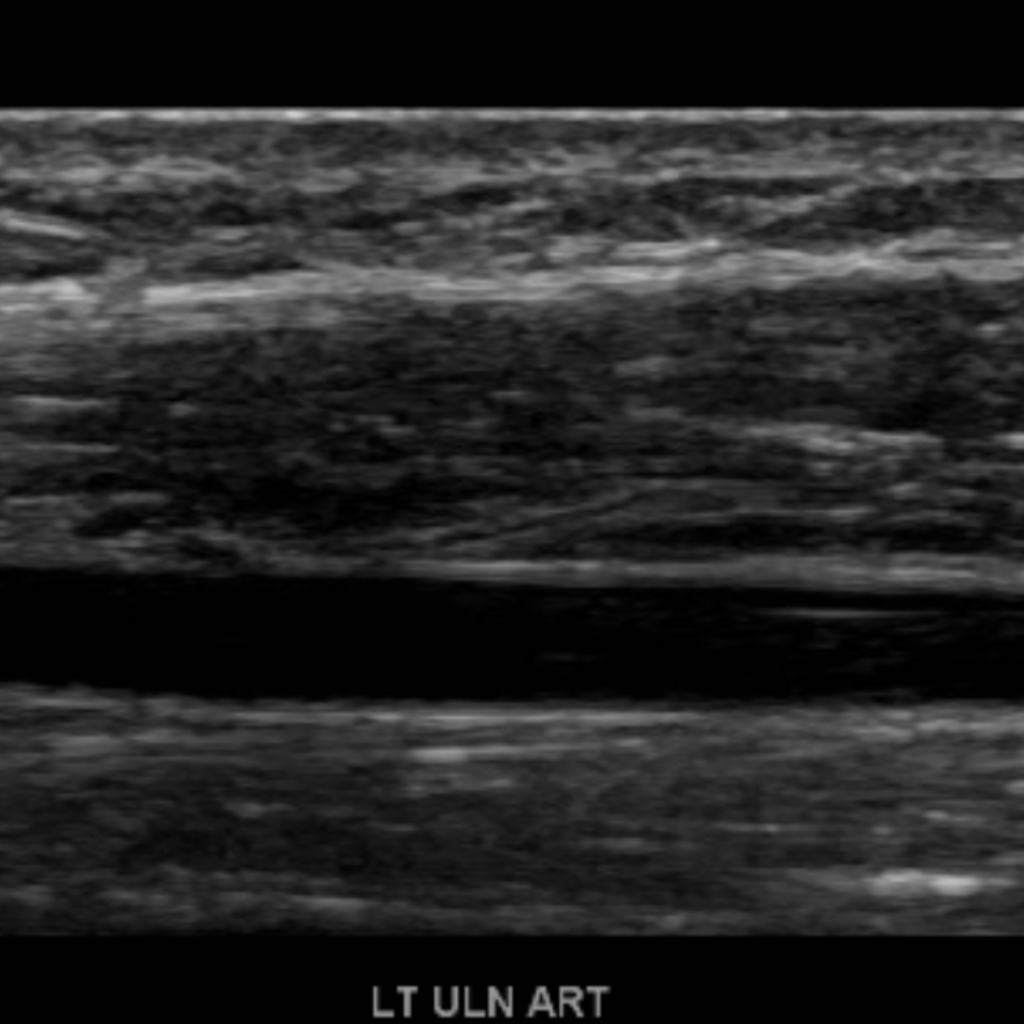
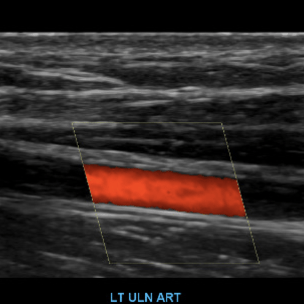
The subclavian artery arises from the brachiocephalic artery on the right and off of the aortic arch on the left. This artery further divides into the axillary, brachial, radial, ulnar, palmar and digital arteries respectively.


[…] Arterial Duplex/Doppler Sonography of the Upper Extremities […]
LikeLike
Absolutely outstanding, thank you, brother!
LikeLike
Thanks!!
LikeLike
Hi Henry, what are the normal velocities of each artery scanned? Subclavian, axiallary, brachial, radial, ulnar? Currently scanning this in school, but have a hard time differentiating and following the vessel.. Was told to look at the velocities to identify each vessel.
LikeLike
Upper extremities anatomy and protocol is very useful. I have a good understanding of the basic knowledge as beginner in Vascular Ultrasound. I am requesting to add more vascular Art/ Vein anatomy and protocol such as, lower extremities, abdominal, and cranial.
LikeLike
[…] August 30, 2025 Arterial Duplex/Doppler Sonography of the Upper Extremities […]
LikeLike